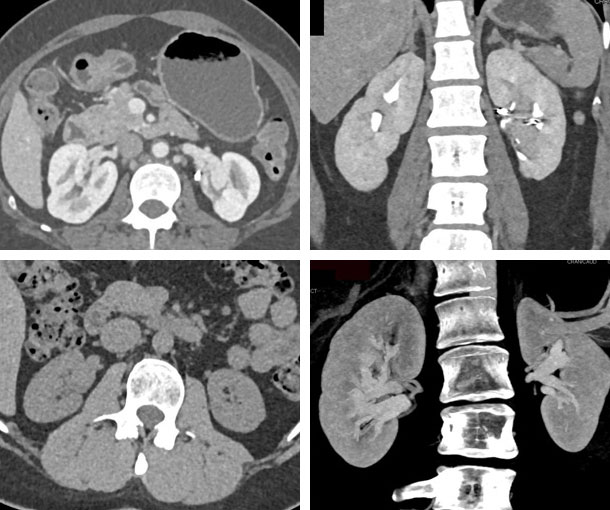
Kidney CT Appearances
Renal AV Malformation (AVM) CT Findings
- Simultaneous opacification of renal arteries and the draining vein
- Best seen typically on arterial phase imaging and poorly seen on excretory phase imaging
- Can be due to trauma (e.g. stab wound, GSW) or be congenital in origin
- Can present clinically with hematuria

Other Information About Renal AVM
Etiology:
- Trauma from renal biopsy or surgery
Epidemiology:
- Peak incidence between 30-40 years of age
- More common in females
Presentation:
- May be asymptomatic
- Flank pain
- Hematuria
- Perinephric hematoma
- Abdominal mass or bruit
- Hypertension
- High-output heart failure
Prognosis:
- Treatment and prognosis depends on how advanced the AVM is
Related Pearls: AVMs
Related Lectures:
CT of the Acute Abdomen: GU Applications Part 3
CTA of the Renal Arteries: What You Need to Know - Part 2
CT of the Aorta & Its Branches: Acute Processes - Part 3
CT Evaluation of Hematuria: A Practical Approach - Part 3
CT of the Abdominal Aorta and Its Branches: Aneurysms, Dissections and Vasculitis - Part 3
Renal Infection Thru Infarction: What You Need to Know - Part 3
