Small Bowel CT Appearances
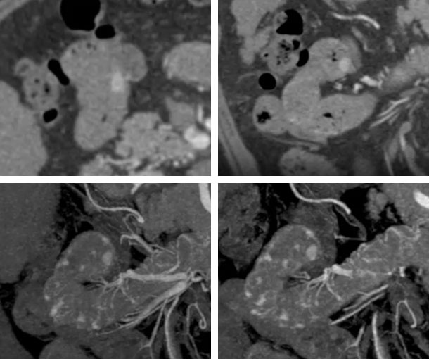
Small Bowel Angiodysplasia CT Findings
- Most common in proximal small bowel but can occur throughout the small bowel
- Tuft-like hypervascular enhancing focus less than 5mm which fades on delayed phases
- Multifocal in 40% to 75% of cases
- Composed of abnormally dilated thin-walled vessels with high propensity for bleeding
Other Information About Small Bowel Angiodysplasia
Etiology:
- Unknown
Epidemiology:
- Typically presents after age 60
Presentation:
- Typically asymptomatic
- Anemia
- Fatigue
- Weakness
- Shortness of breath
Prognosis:
- For symptomatic patients, treatment can successfully control the bleeding
Related Lectures:
CT Evaluation of GI Bleeding - Part 3
CT Evaluation of GI Bleeding - Part 4
Abdominal Pain in the ER: GI Pathology - Part 3
CT Evaluation of GI Bleeding - Part 2
CT Evaluation of GI Bleeding - Part 3