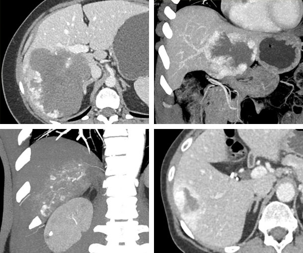
Liver CT Appearances
Hepatic Hemangioma CT Findings
- Lesion with large vascular spaces and a central cavernous area
- Well defined outline with peripheral enhancement
- Homogenous on delayed phase imaging
- “Giant Cavernous Hemangioma” is defined as lesions > 3cm
- Over 90% of hemangiomas have the classic peripheral puddling appearance

Other Information About Hepatic Hemangioma
Etiology:
- Unknown
Epidemiology:
- More common in females
- Patients usually present at age 30-50
Presentation:
- Asymptomatic
Prognosis:
- Hepatic hemangiomas are benign
Related Pearls: Hemangioma
Related Lectures:
CT Evaluation of Liver Masses: Key Differential Diagnosis Findings - Part 2
CT Evaluation of Liver Masses: Key Differential Diagnosis Pathways - Part 2
CTA 2024: Oncologic Applications in Clinical Practice - Part 1
CT Evaluation of Hepatic Masses: A Focused Approach to Signs and Enhancement Patterns - Part 3
