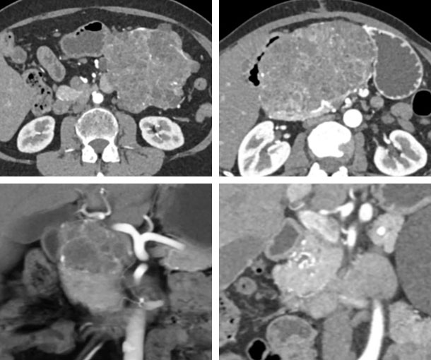
Pancreas CT Appearances
Serous Cystadenoma CT Findings
- Calcifications in up to 30% of cases
- Patterns of calcification are variable
- More common in body and tail of pancreas
- Classic cases are easy to diagnose but many are atypical
- Multiple cysts with thin septations
- Central scar with central calcification is classic
- Less common appearance is oligocystic variety (10%) with single cyst and hard to distinguish from MCN
- Cysts contain glycogen but no mucin
- Polycystic pattern is most common in 70% of cases and cysts measure 2cm or smaller
- Honeycomb pattern is second most common in 20% of cases and numerous cysts under 1cm in size
- Numerous cysts can typically not be separated individually
- Oligocystic is least common in less than 10% of cases and a few cysts over 2cm in size
- Commonly called macrocystic cystadenoma and can be confused with mucinous cystic tumors

Other Information About Serous Cystadenoma
Etiology:
- Unknown
- Associated with von Hippel-Lindau
Epidemiology:
- Typically present in 5th to 7th decades of life
Presentation:
- Often asymptomatic
- Abdominal pain
- Palpable mass
Prognosis:
- Serous cystadenomas are benign
Related Lectures:
Cystic Pancreatic Lesions: What You Need to Know - Part 1
CT of Serous Cystadenomas of the Pancreas: A Lesion of Many Different Appearances - Part 1 CT of Serous Cystadenomas of the Pancreas: A Lesion of Many Different Appearances - Part 2
CT of Serous Cystadenomas of the Pancreas: A Lesion of Many Different Appearances - Part 3
Cystic Pancreatic Lesions: Detection, Diagnosis and Management - Part 3
CTA 2024: Oncologic Applications in Clinical Practice - Part 2
Learning Modules:
Characterization of Pancreatic Serous Cystadenoma on Dual-Phase Multidetector CT
