Stomach CT Appearances
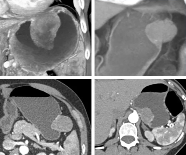
Gastric GIST Tumor CT Findings
Benign
- Well-circumscribed endophytic, exophytic, or bilobed mass with its epicenter in the submucosa
- Ulceration can be visualized in lesions larger than 2cm (bullseye sign)
Aggressive
- Large (>5cm) heterogenous mass
- Necrosis, hemorrhage, with or without calcifications
- Lymphadenopathy, with or without metastases
- Invasion of adjacent viscera (pancreas, colon)
- Ulcerations very common in malignant GISTs
- Malignant GISTs are often inhomogenous with central necrosis
- Metastases to liver is most common site of metastases
Other Information About Gastric GIST Tumors
Etiology:
- Unknown
- Risk increases with familial history
- Linked to NF1 and Carney-Stratakis syndrome
Epidemiology:
- Typically present after age 50
Presentation:
- Typically asymptomatic
- Abdominal pain
- Palpable mass
- Fatigue
- Anorexia
- Weight loss
- Hematochezia
Prognosis:
- The 5-year relative survival rates for all GISTs are:
- Localized: 95%
- Regional: 84%
- Distant: 52%
- All SEER stages combined: 85%
Related Pearls: GIST Tumors
Related Lectures:
CT Evaluation of Gastric Tumors: Pearls and Pitfalls Part 2
MDCT Evaluation of Gastric Malignancies: Pearls and Pitfalls - Part 2
Gastric GIST Tumors: Pearls and Pitfalls - Part 1
Gastric GIST Tumors: Pearls and Pitfalls - Part 2
Gastric GIST Tumors: Pearls and Pitfalls - Part 3
CT of the Stomach: Beyond the Common Gastric Masses - Part 1