Spleen CT Appearances
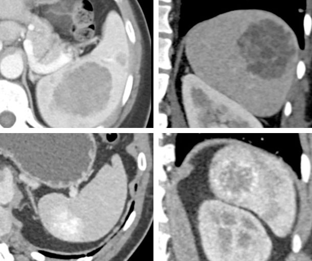
Splenic Hemangioma CT Findings
- May enhance similar to hepatic hemangiomas
- May remain hypodense or hyperdense
- May have calcifications
- Can appear solid or cystic
Other Information About Splenic Hemangioma
Etiology:
- Unknown
Epidemiology:
- Rare
- Typically presents in 4th-6th decades of life
Presentation:
- Typically asymptomatic
- May rupture which would cause abdominal pain (rare)
Prognosis:
- Symptomatic patients may have a splenectomy
Related Lectures:
MDCT Evaluation of the Spleen: Challenges in Diagnosis Part 2
Challenging Cases in CT: A Case Based Approach to the Incidental Splenic Mass - Part 2
CT Evaluation of Incidentalomas: A Practical Set of Strategies - Part 4
MDCT Evaluation of the Spleen: Challenges in Diagnosis - Part 2
MDCT Evaluation of the Spleen: Challenges in Diagnosis - Part 3