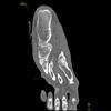
Foot Pathology: Talar Fractures
The talus is the second most common tarsal bone to fracture. The paucity of muscular attachments makes this bone prone to dislocation following trauma. The most common fractures are osteochondral fractures and avulsion fractures.
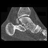
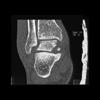
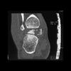
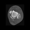
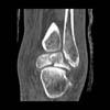
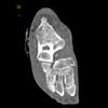
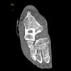
Osteochondral lesions of the talar dome can be challenging to detect with conventional radiographs, and carry a worse prognosis compared to other avulsion fractures. Failure to detect this lesion can result in prolonged disability. In the presence of an ankle joint effusion with no fracture apparent on convential radiograph, CT should be performed if there is a high clinical suspicion for this type of lesion. The most common cause is trauma. Other hypothesized etiologies include ischemic necrosis, spontaneous necrosis, abnormal vascular anatomy and congenital abnormalities.
Osteochondral lesions of the talus are much more common in men (72%). These lesions can occur medially or laterally in the talar dome. The medial lesions occur in the posterosuperior half of the dome of the talus; however, the lateral lesions are situated anterosuperiorly. The lateral lesions almost always follow trauma; however, a history of trauma is presented in only 70% of those with medial lesions.
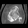
MR is helpful to assess the integrity of the articular cartilage and to detect loose intraarticular bone fragments. However, complete assessment often requires arthroscopy or fluoroscopy.
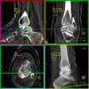
Treatment depends on the stage of the lesion, as defined by Berndt and Harty. Stage I is defined as a small region of subchondral bone compression. Stage II fractures are incomplete, with partial detachment of the osteochondral fragment. Stage III includes all completely detached but nondisplaced fractures. A displaced, detached fragment constitutes a Stage IV lesion. Stage I-III lesions can be treated by drilling through the sclerotic margin of the lesion in increase perfusion. Intraoperative fluoroscopy has traditionally been used to guide this treatment; however, the utility of computer assisted navigation has recently been reported for preoperative planning.
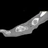
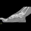
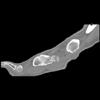
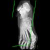
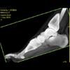
Fractures of the talar neck are seen in the setting of motor vehicle accidents, falls or direct trauma, as well as other traumatic events that induce an abrupt force from below and dorsiflex the foot. Talar neck fractures are classified by the presence and degree of vertical dislocation. Stage I includes nondisplaced vertical neck fractures. The presence of subtalar dislocation or subluxation equates with a stage II fracture, which usually disrupts 2-3 of the sources of blood supply to the bone. Stages III includes those fractures with subtalar and tibiotalar subluxation or dislocation, and stage IV fractures are vertically displaced with subtalar or tibiotalar subluxation or dislocation, as well as talonavicular subluxation or dislocation. These latter types of injury generally disrupt all 3 sources of blood supply to the talus. Displaced fractures are commonly complicated by ischemia as a result of the blood supply disruption. CT is necessary for proper delineation of the extent of a talar fracture. In addition, CT is very useful for follow up to determine joint alignment, to exclude any residual intrarticular bone fragments and to detect complications of nonunion and osteoarthritis.

