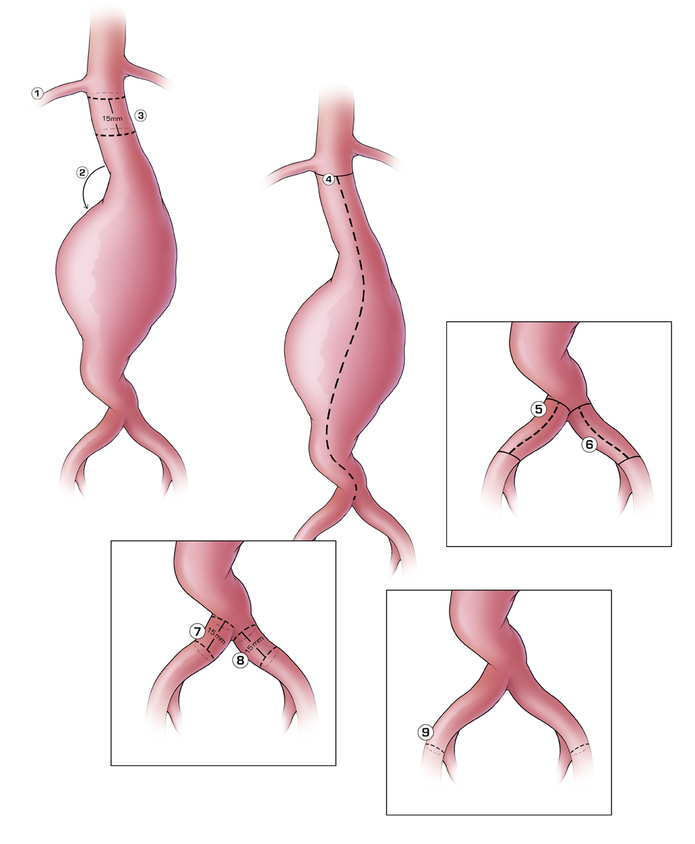
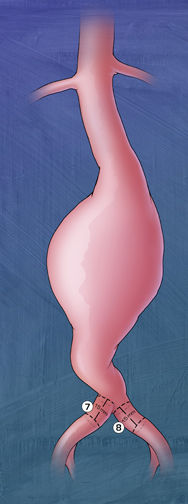
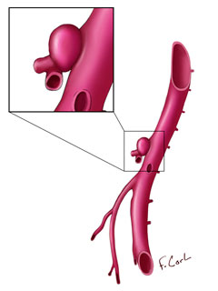
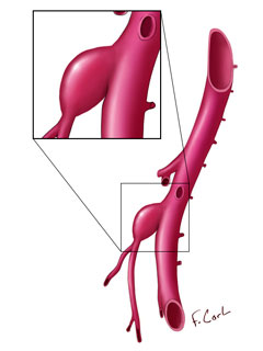
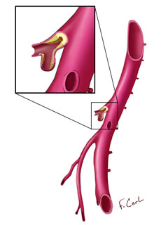
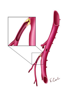
Abdominal Aorta Stent Measurements
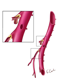
Medical Illustrations showing the important measurements, placement, and angles to consider when planning for stent placement in the abdominal aorta.
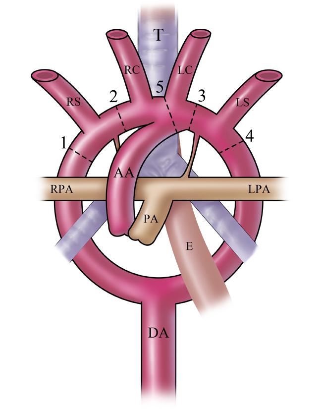
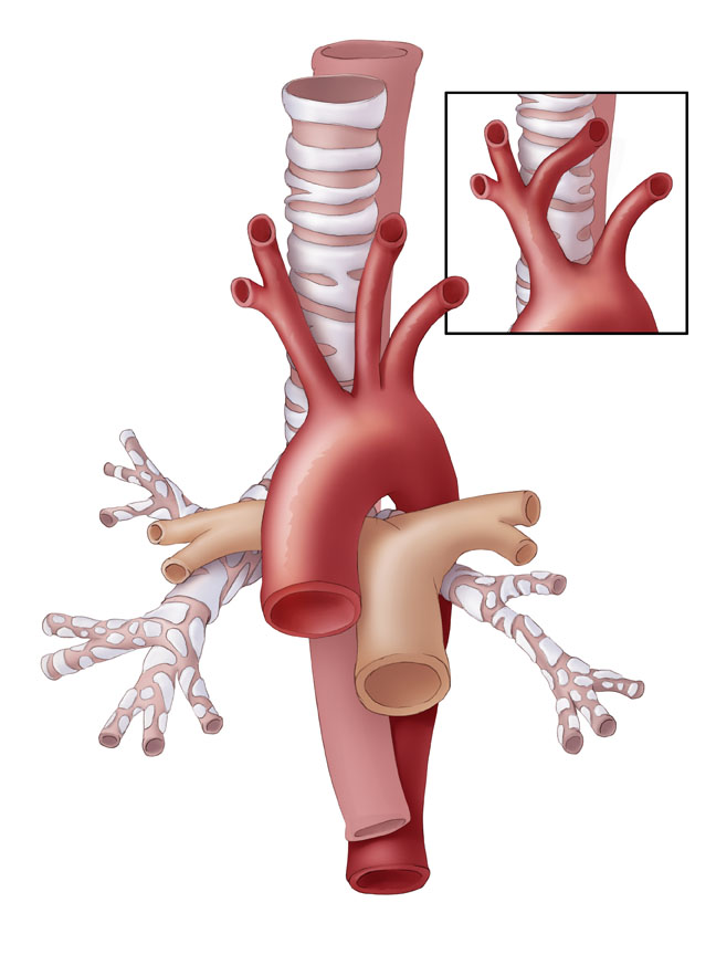
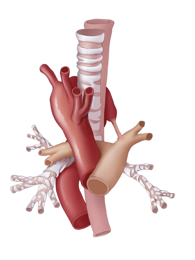
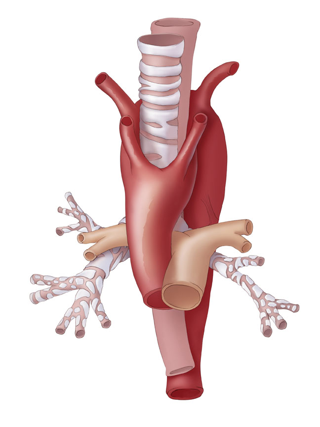
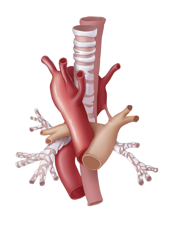
Aortic Arch Anatomy:
Common and Unusual Variants
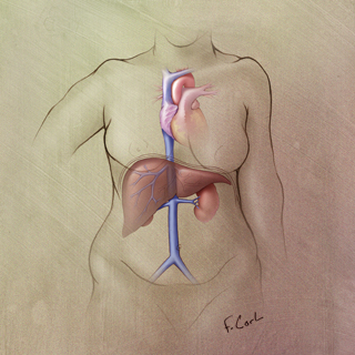
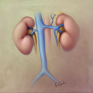
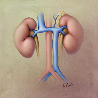
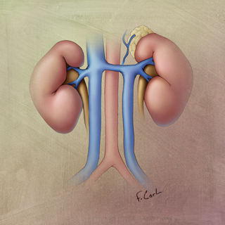
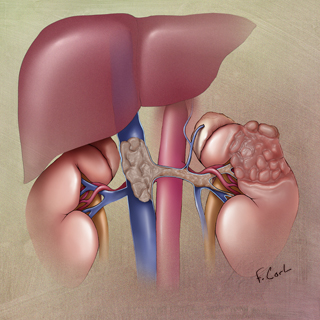
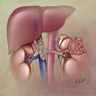
Evaluation of the Inferior Vena Cava
MDCT Evaluation of the Inferior Vena Cava (IVC): Spectrum of Disease with a Focus on Genitourinary Tract Involvement of the IVC
Sheth S, Horton KH, Fishman, EK
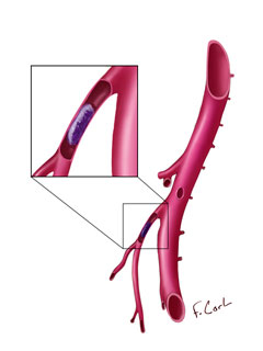
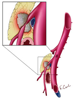
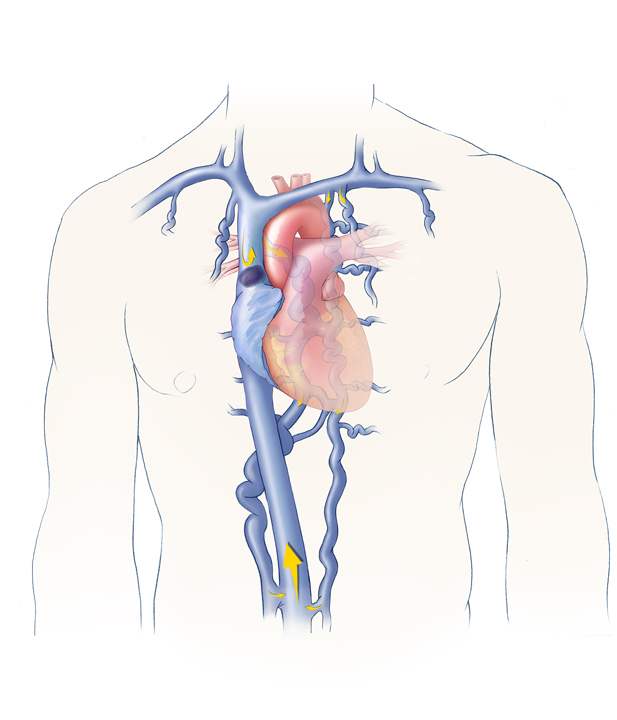
These medical illustrations depict specific pathological conditions and processes of the IVC, such as anatomic variation and malignancy. These illustrations were part of RSNA exhibit and journal publication demonstrating the role of MDCT in the diagnosis of disease processes affecting the IVC.
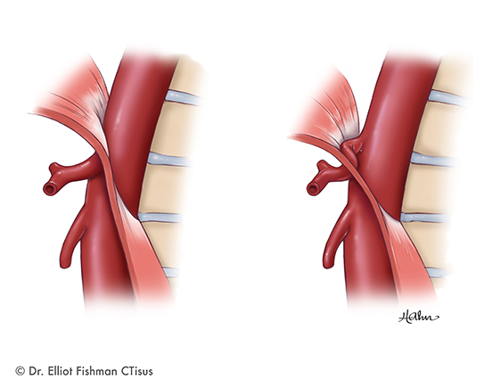
Median Arcruate Ligament Syndrome
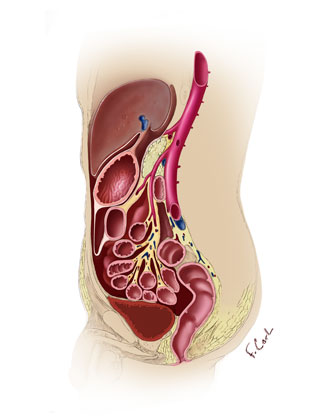
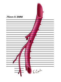
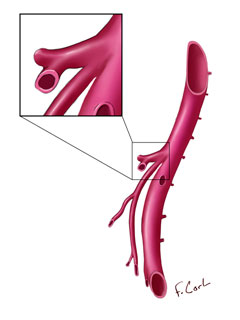
Pathology of the Superior Mesenteric Artery
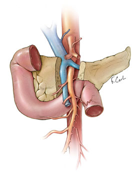
These medical illustrations show the superior mesenteric artery (SMA) and surrounding anatomy, specific types of pathology, scanning protocols, and variant anatomy.
Preoperative Breast Reconstruction Planning
SMA and Celiac Artery Anatomy
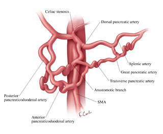
These medical illustrations depict the normal celiac and SMA vascular anatomy and SMA/celiac collaterals due to celiac stenosis.
Subclavian Arterial Mapping
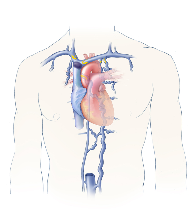
SVC
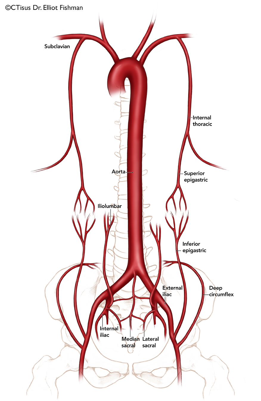
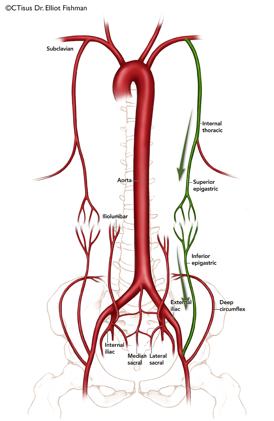
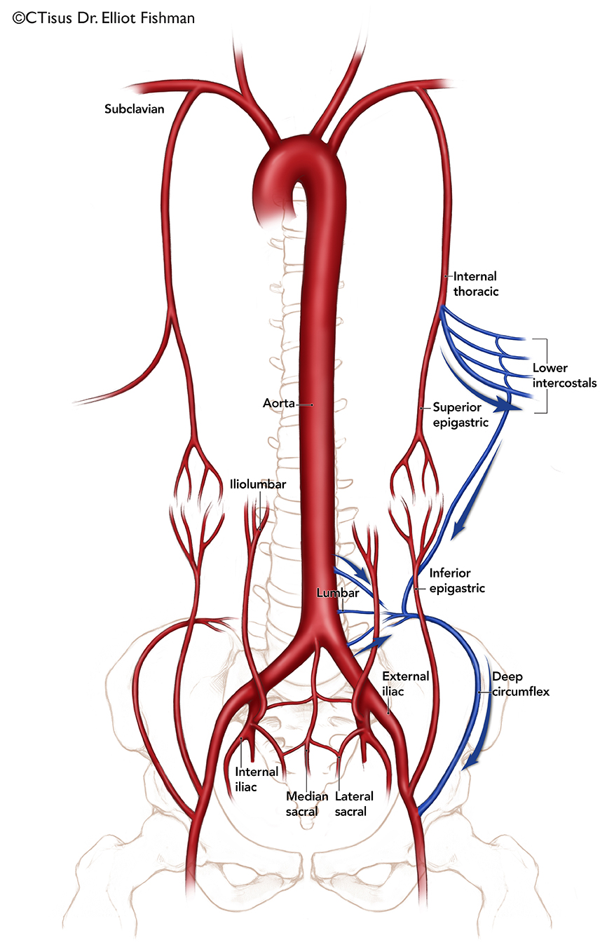
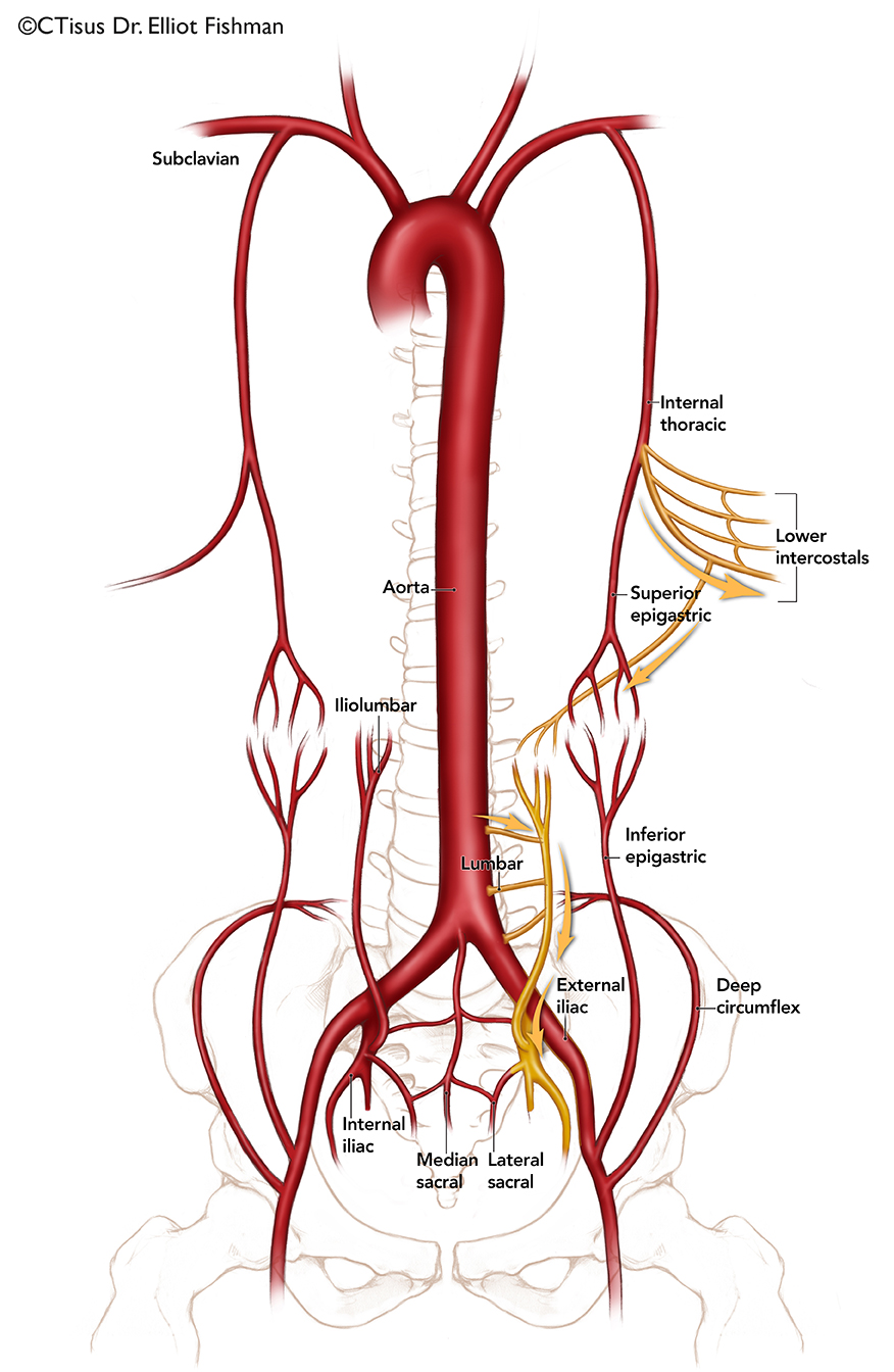
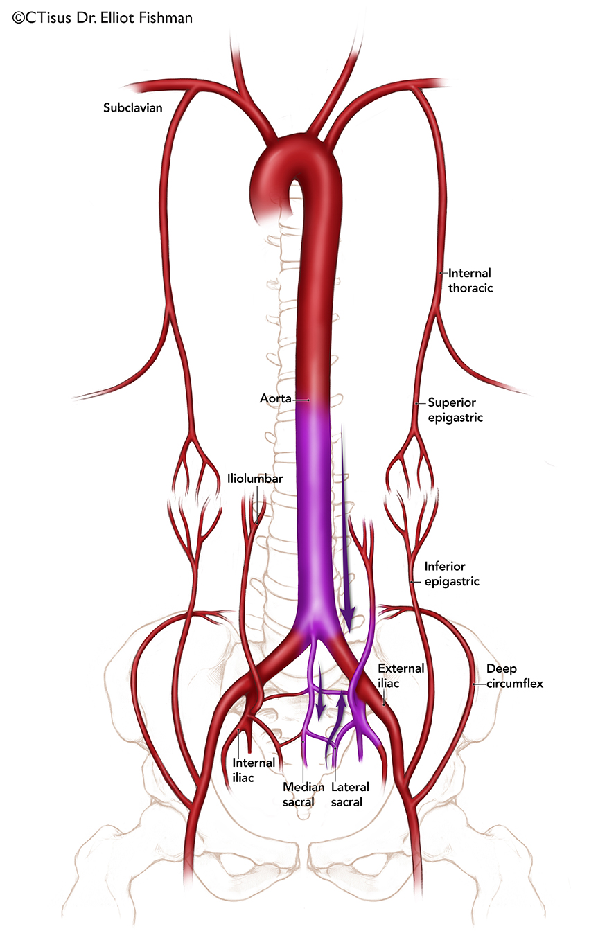
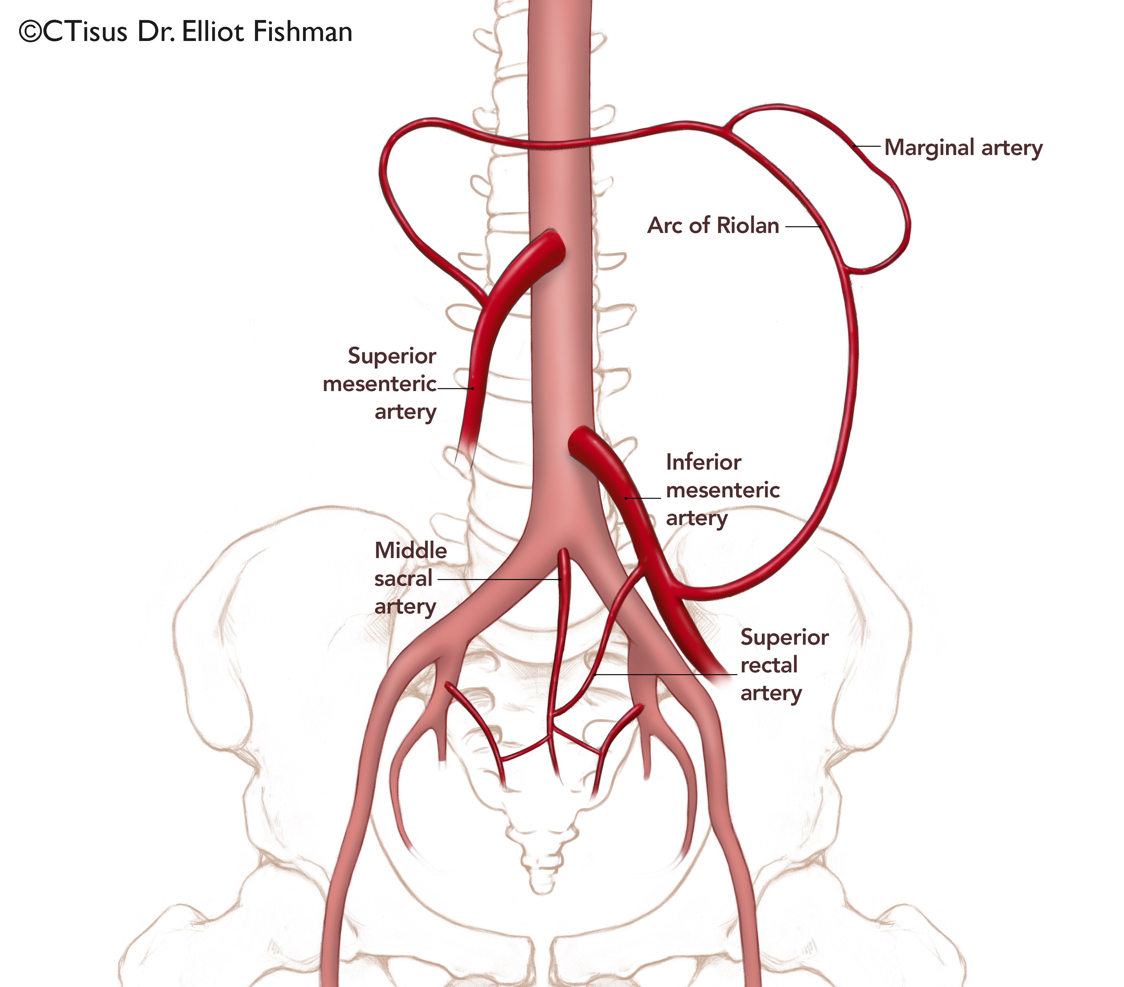
Systemic and Visceral Collaterals
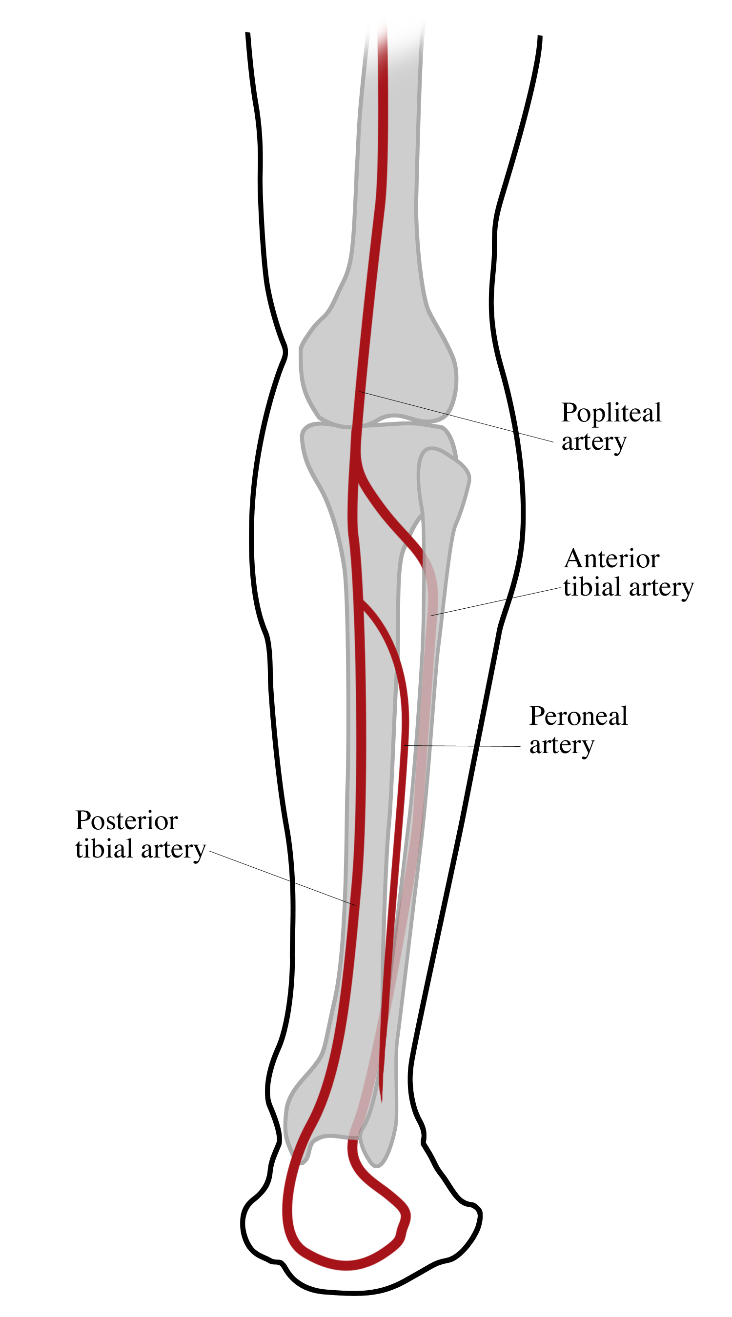
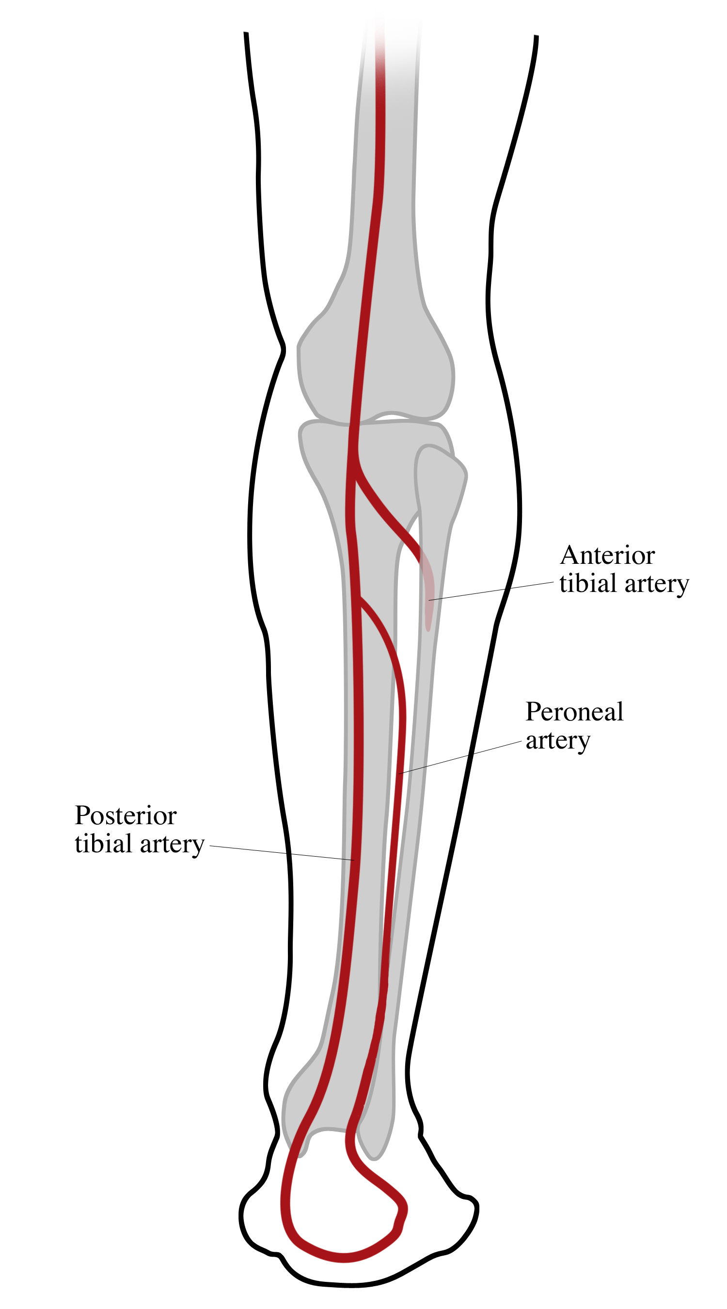
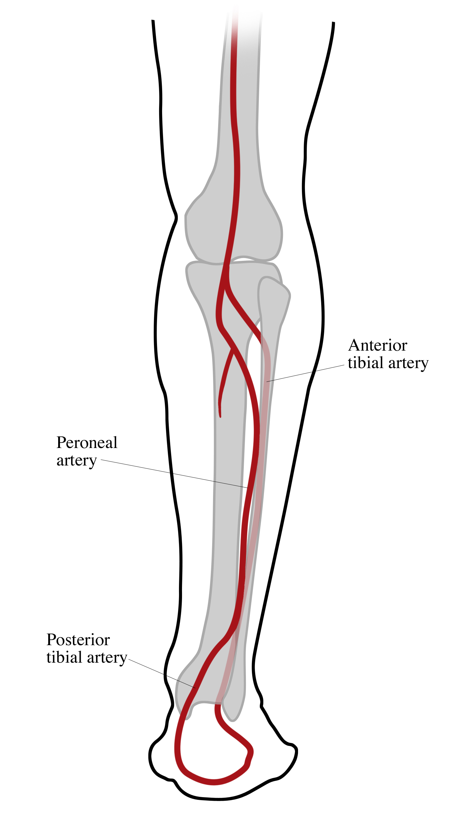
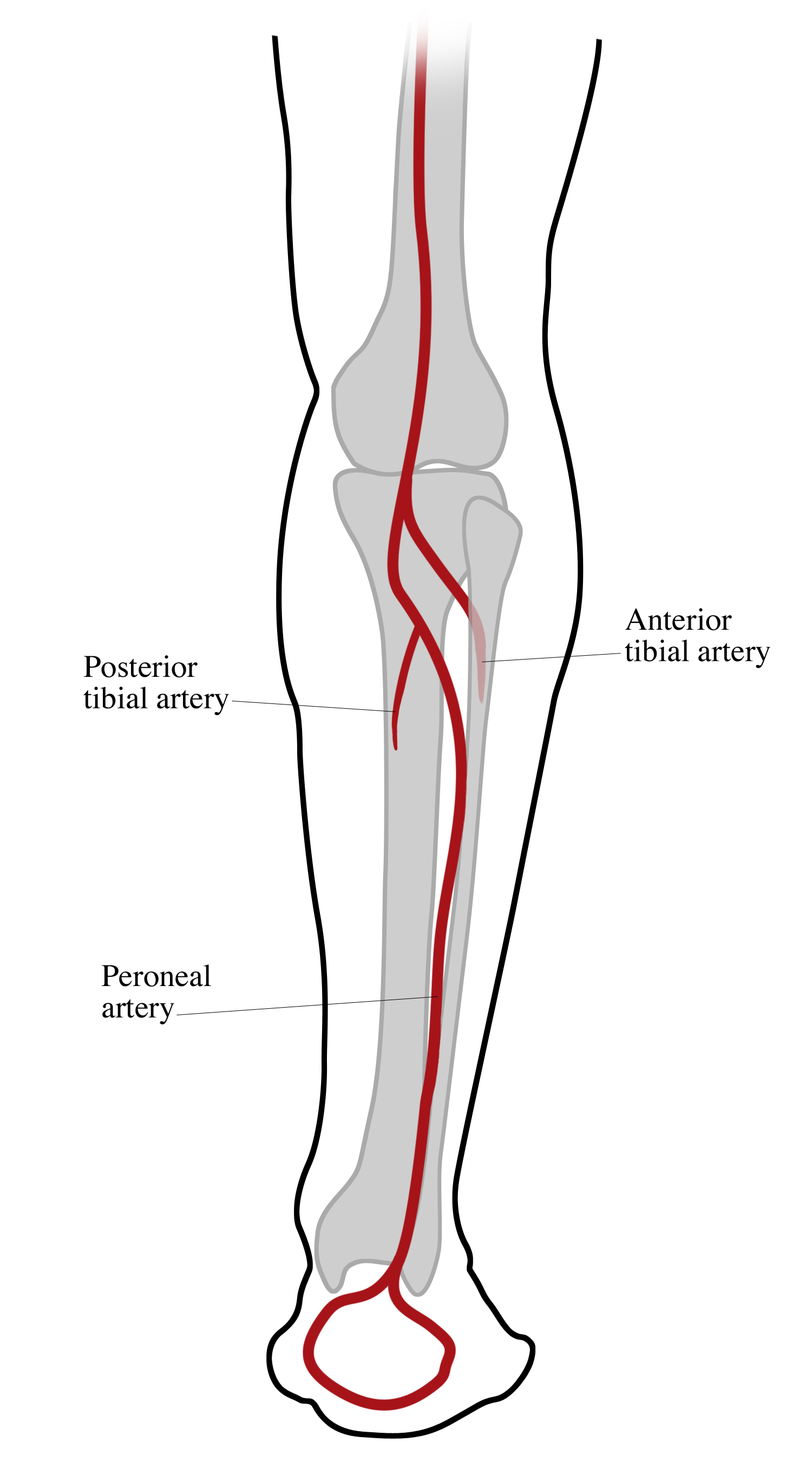
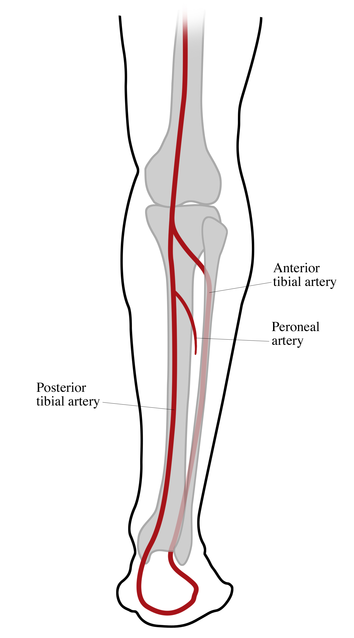
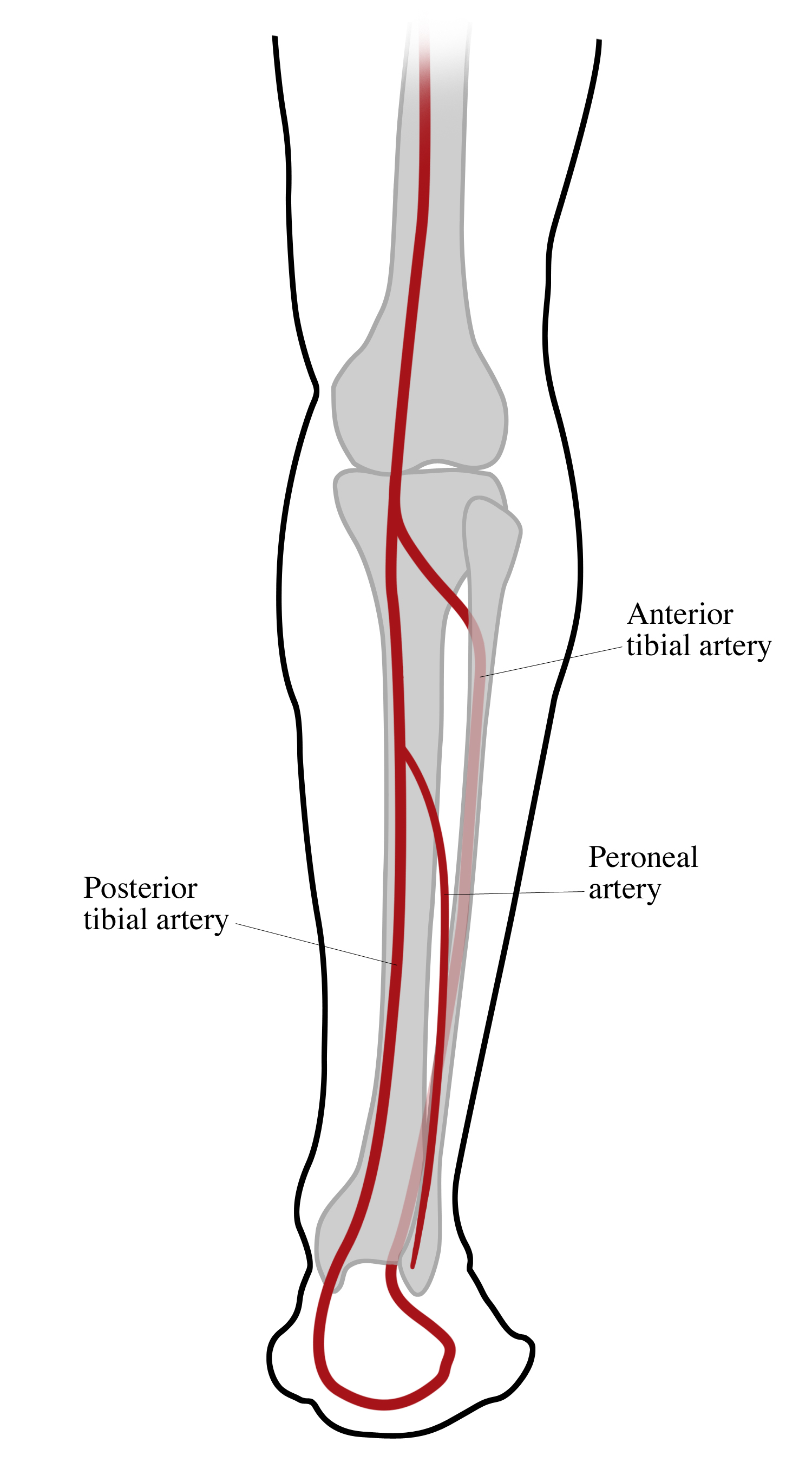
Tibial Artery
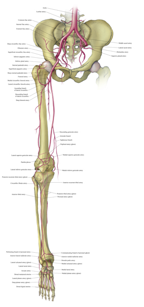
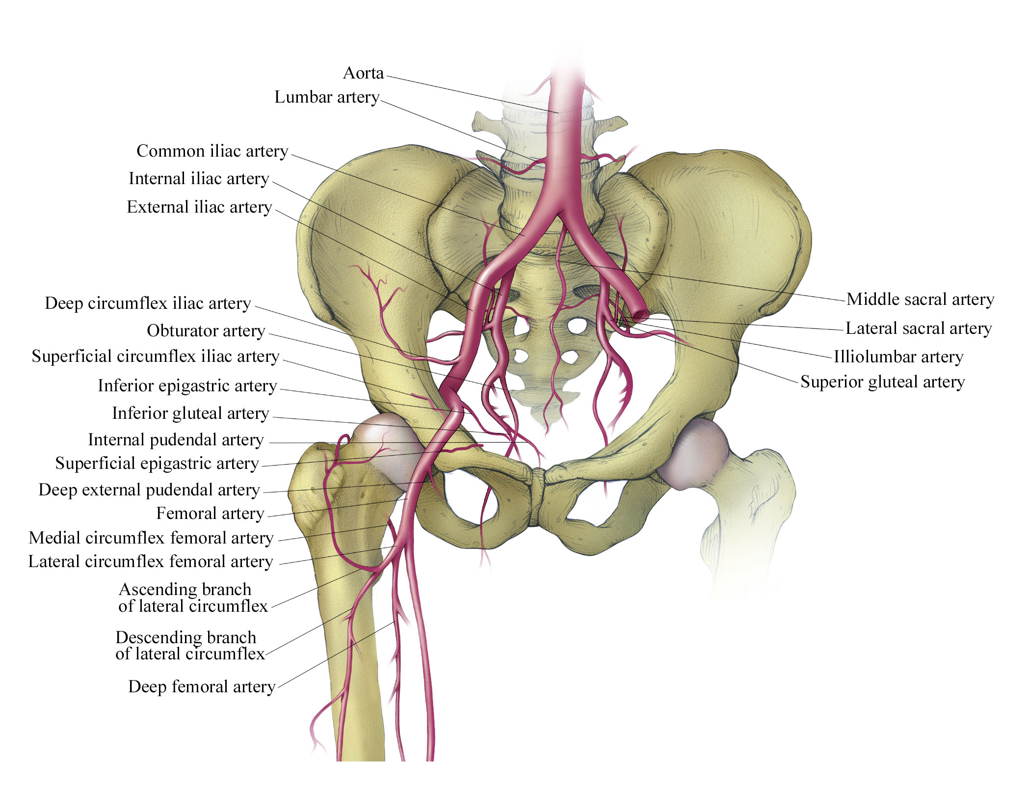
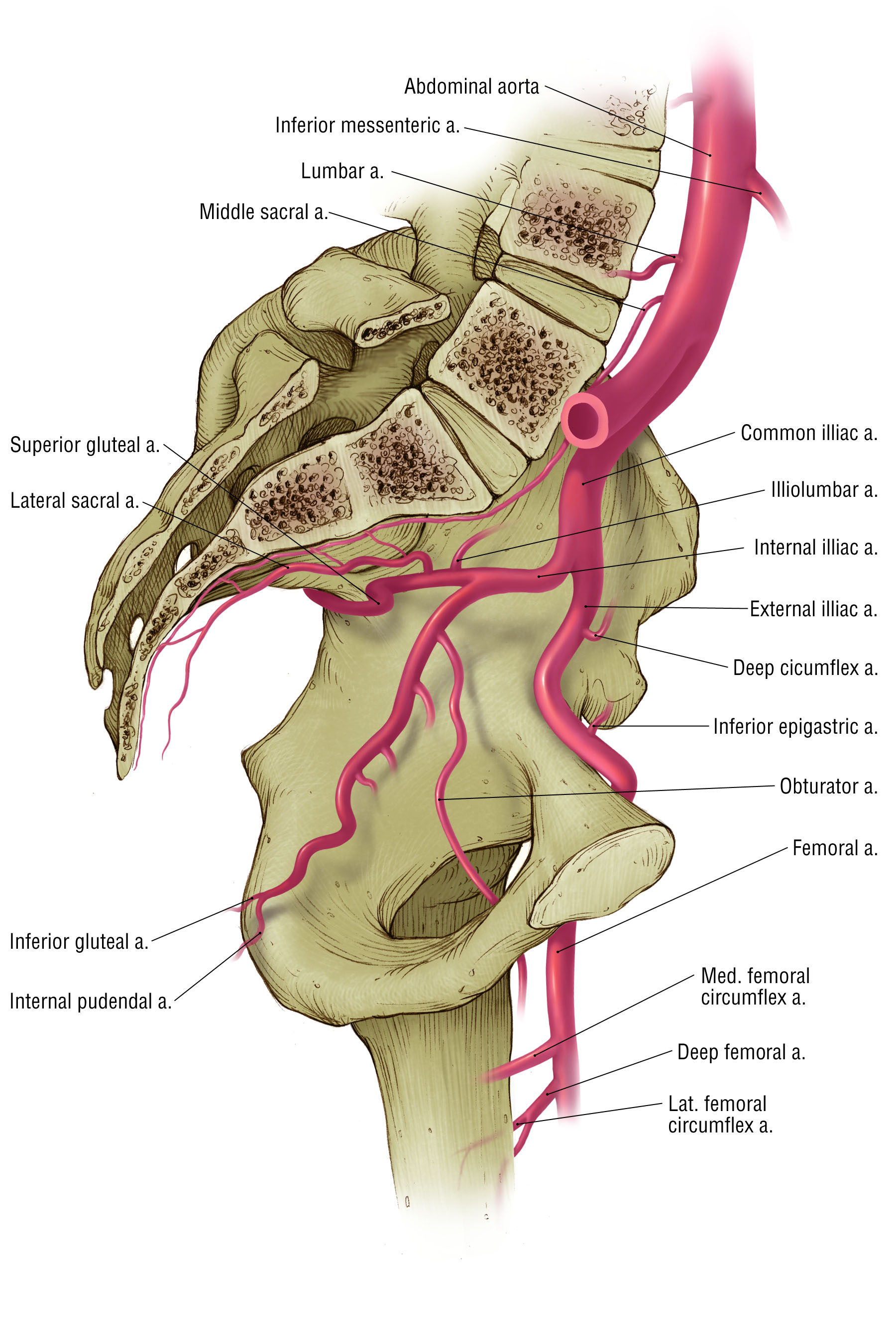
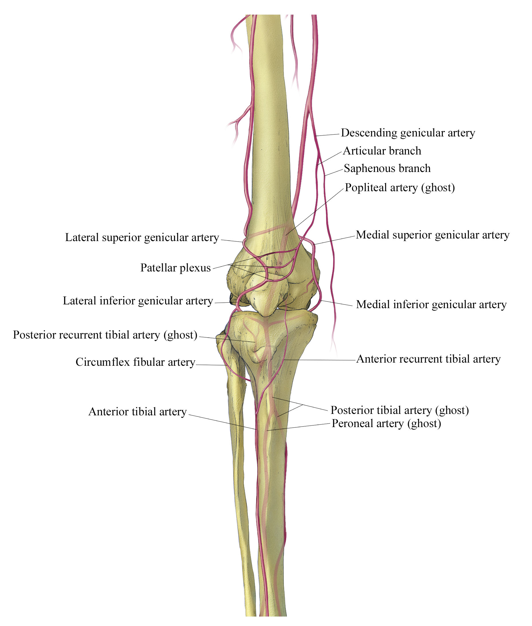
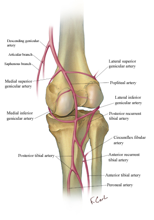
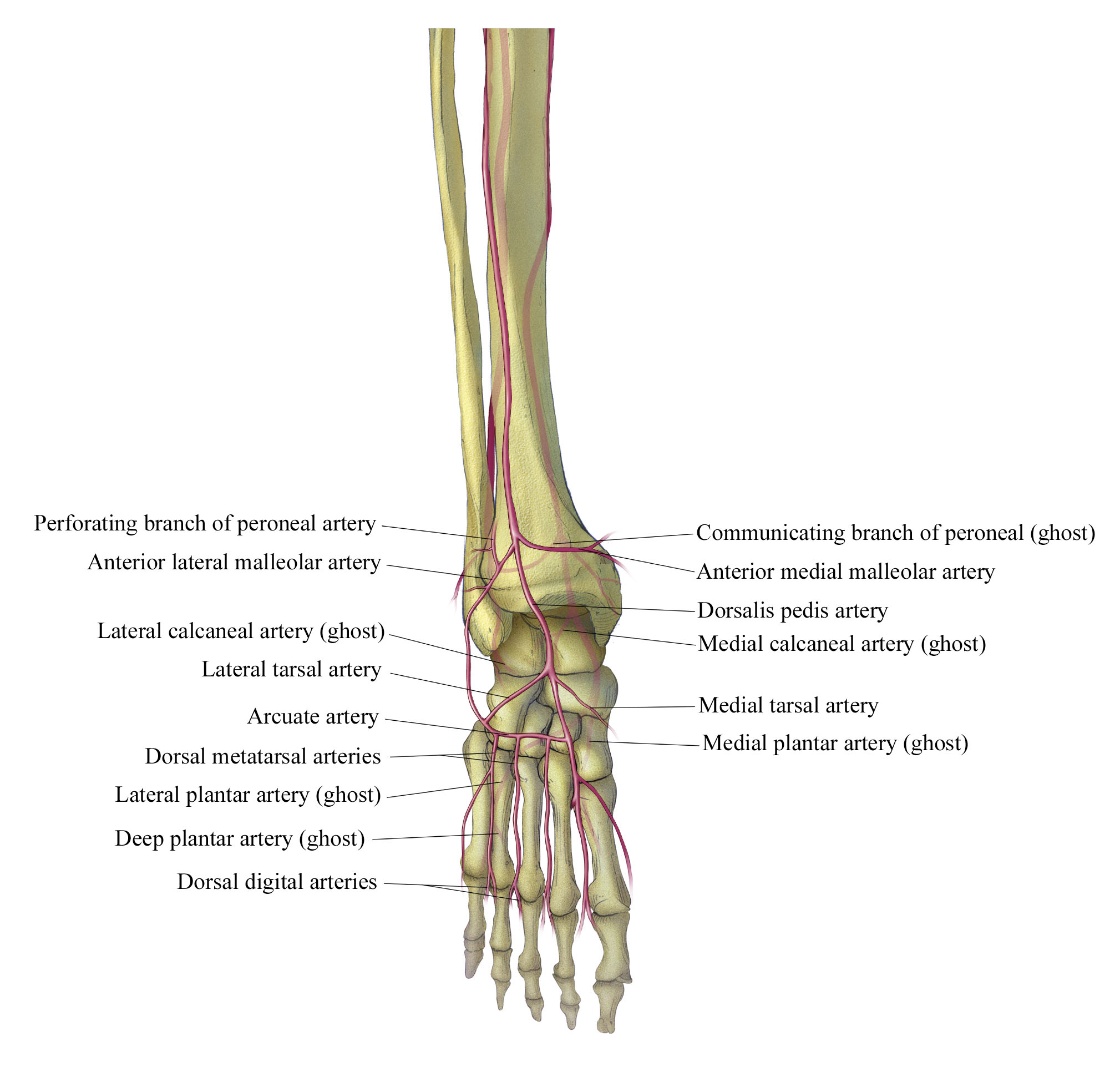
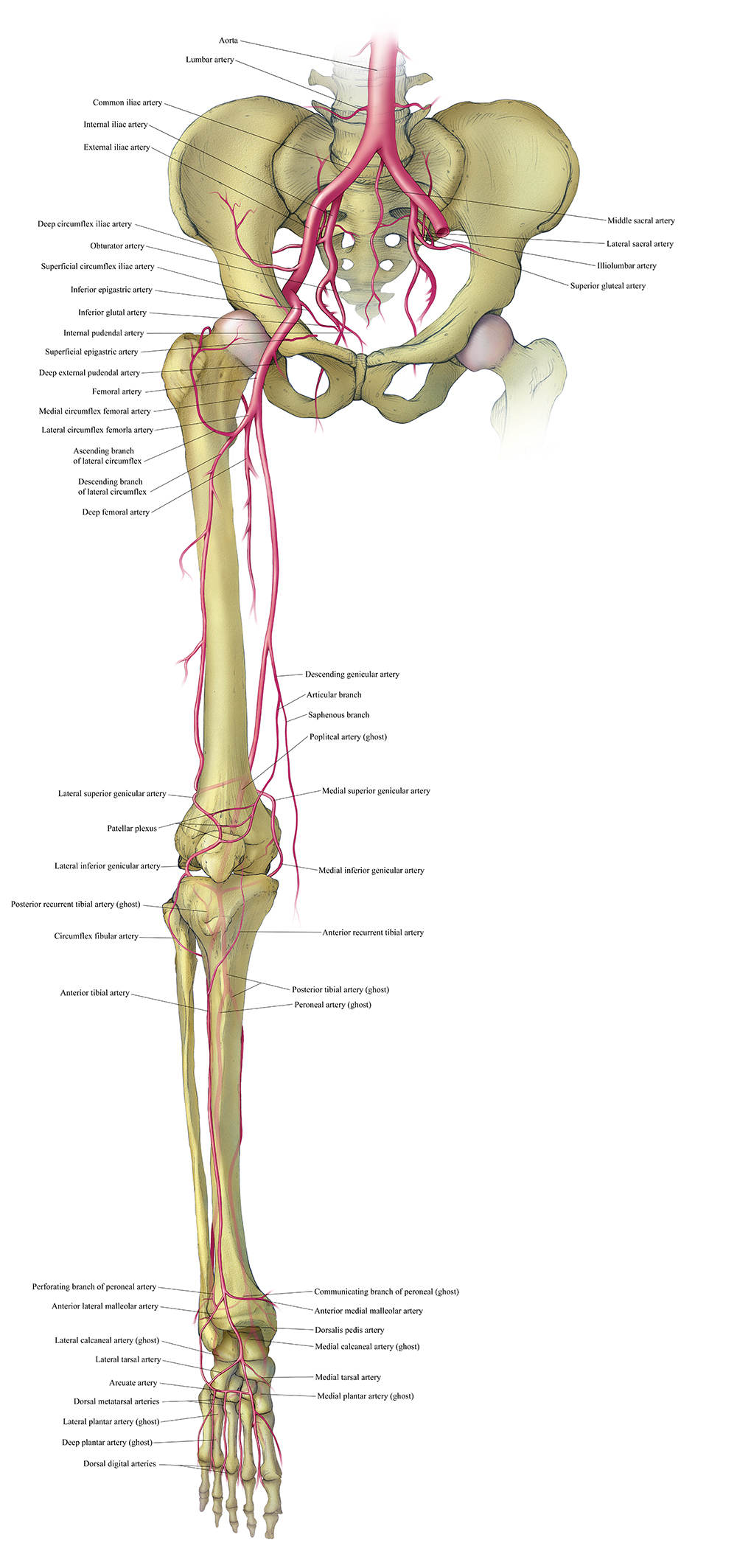
Vasculature of the Lower Extremity
These medical illustrations show normal vascular anatomy of the lower extremity
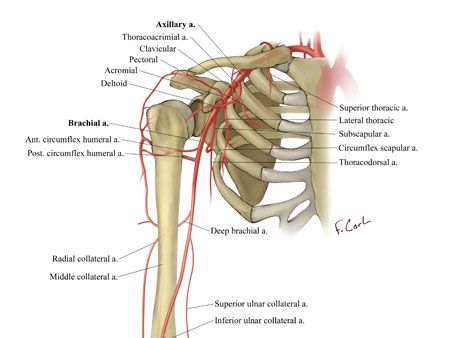
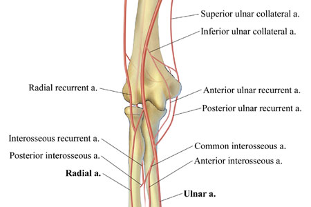
Vasculature of the Upper Extremity
Evaluation of the Utility of Upper Extremity CT Angiography Using 16 and 64 Slice Multidetector CT with Three- Dimensional Volume Rendering
Fishman EK, Smith LS, Neyman EG, Corl F, Lawler LP
These medical illustrations were part of an RSNA exhibit that discussed normal vascular anatomy of the upper extremity, the utility of multidetector CTA (MDCTA) in demonstrating this anatomy, CT protocols for performing CTA of the upper extremity, and a wide variety of cases.

