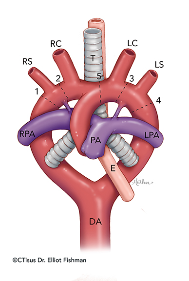
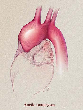
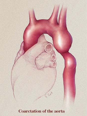
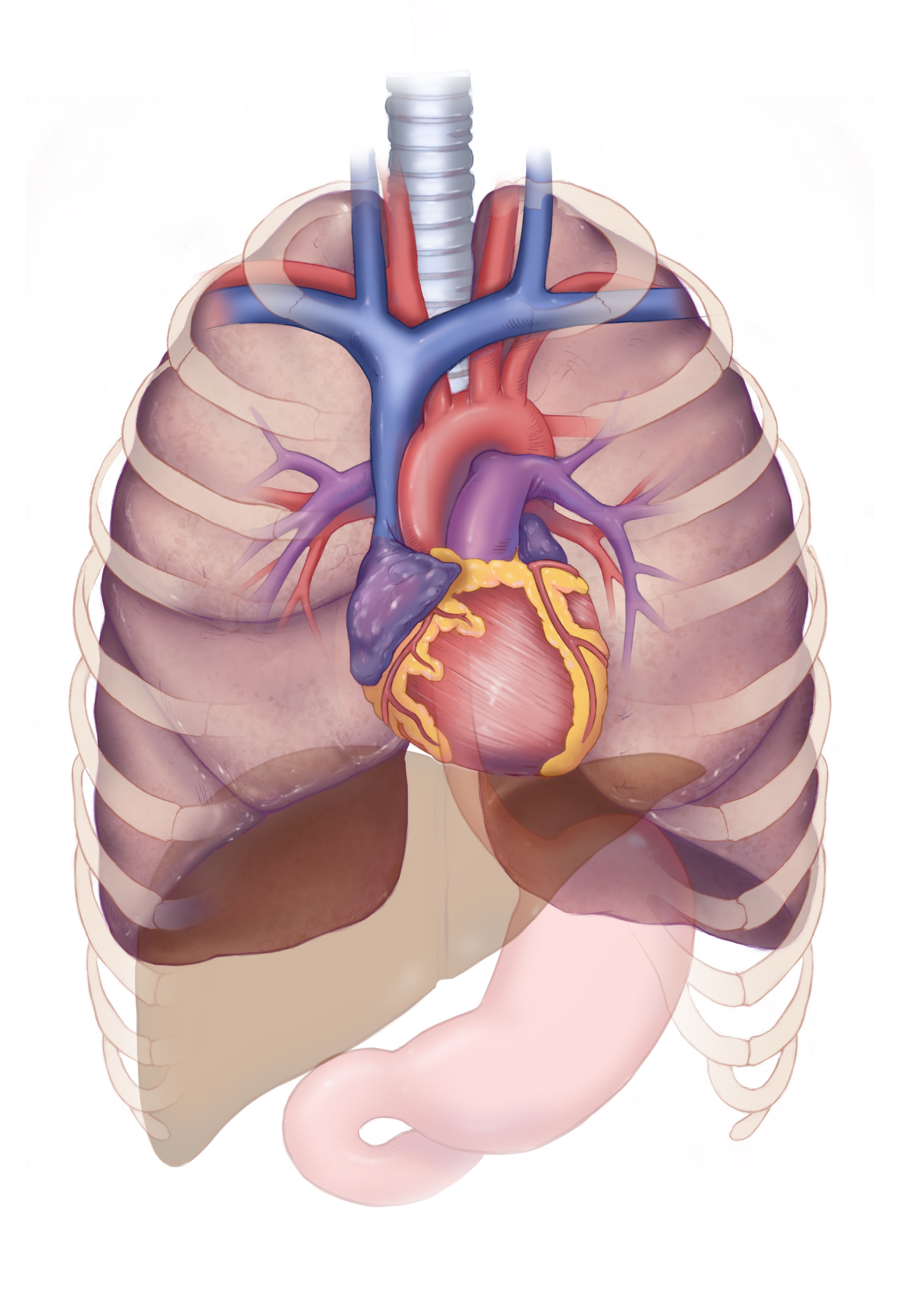
Aortic Aneurysm
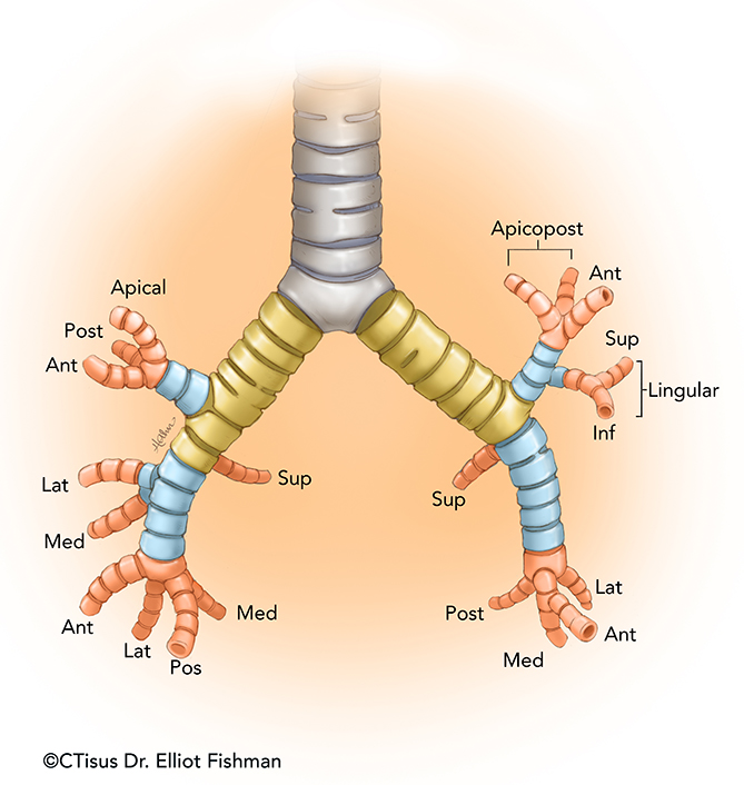
Bronchus Segments
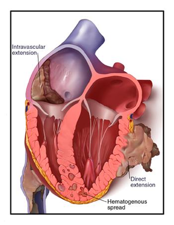
Cardiac Tumors
Embryological Heart
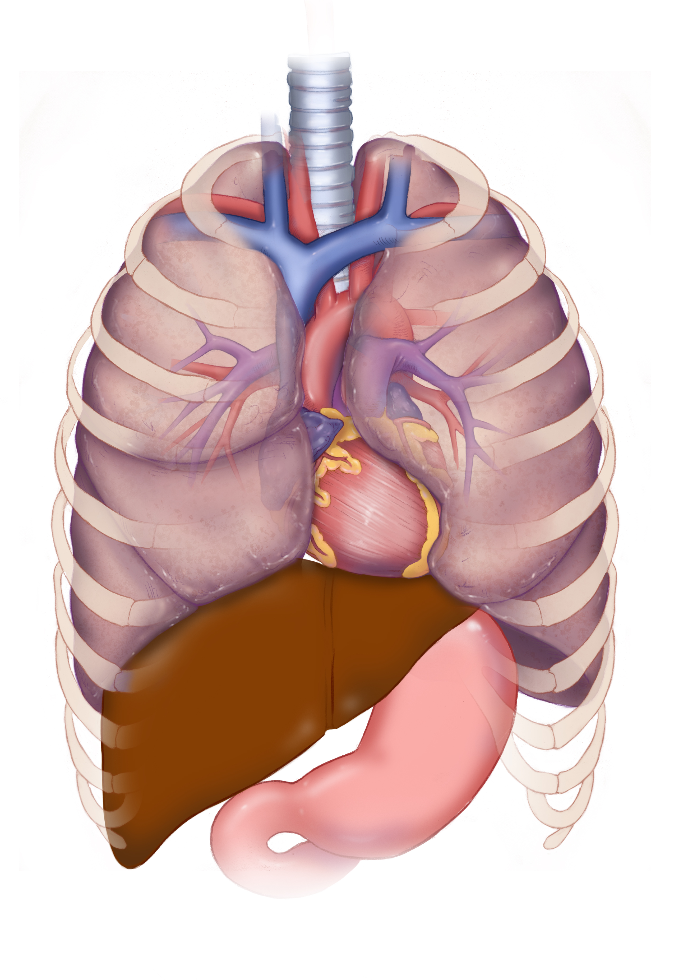
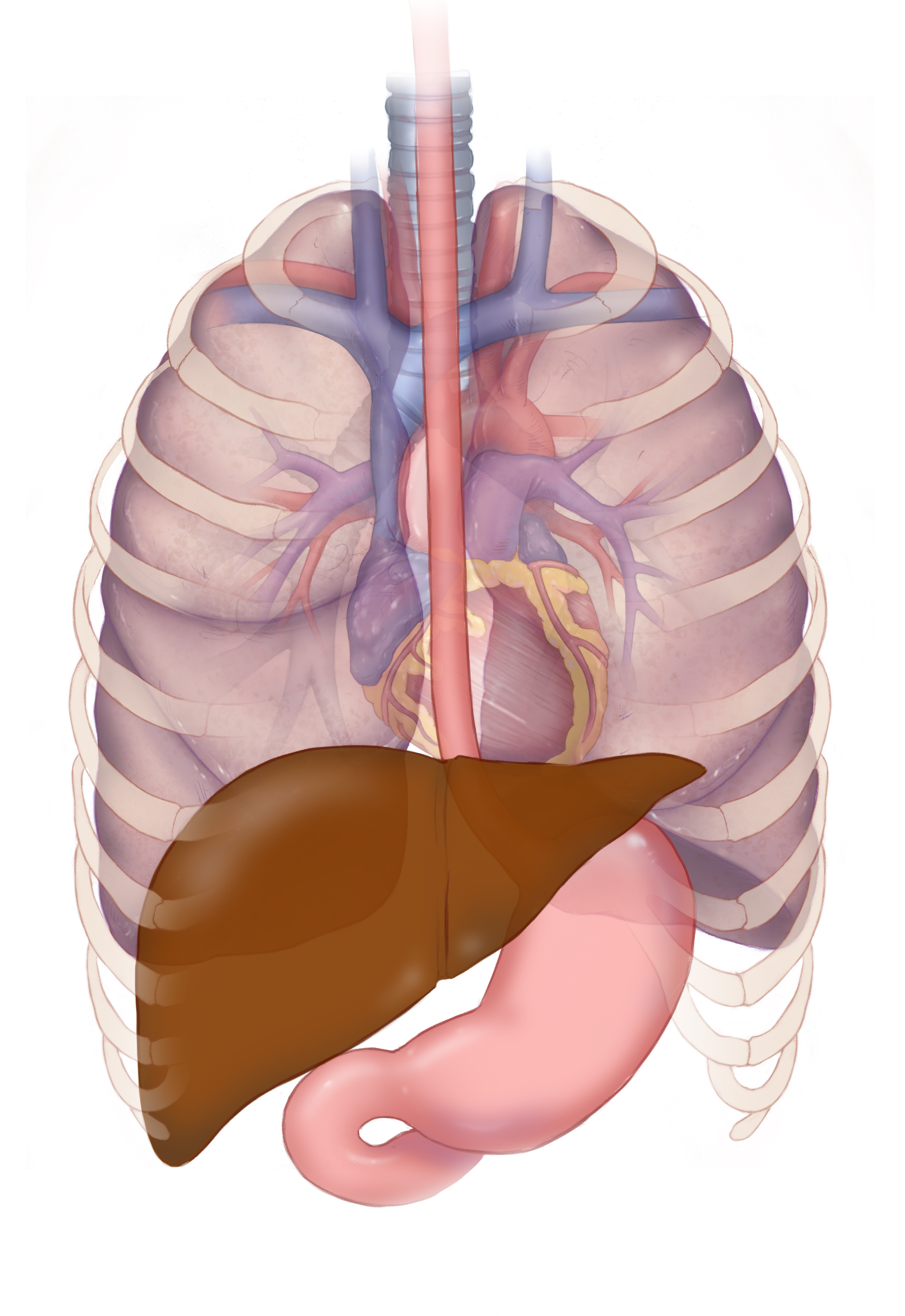
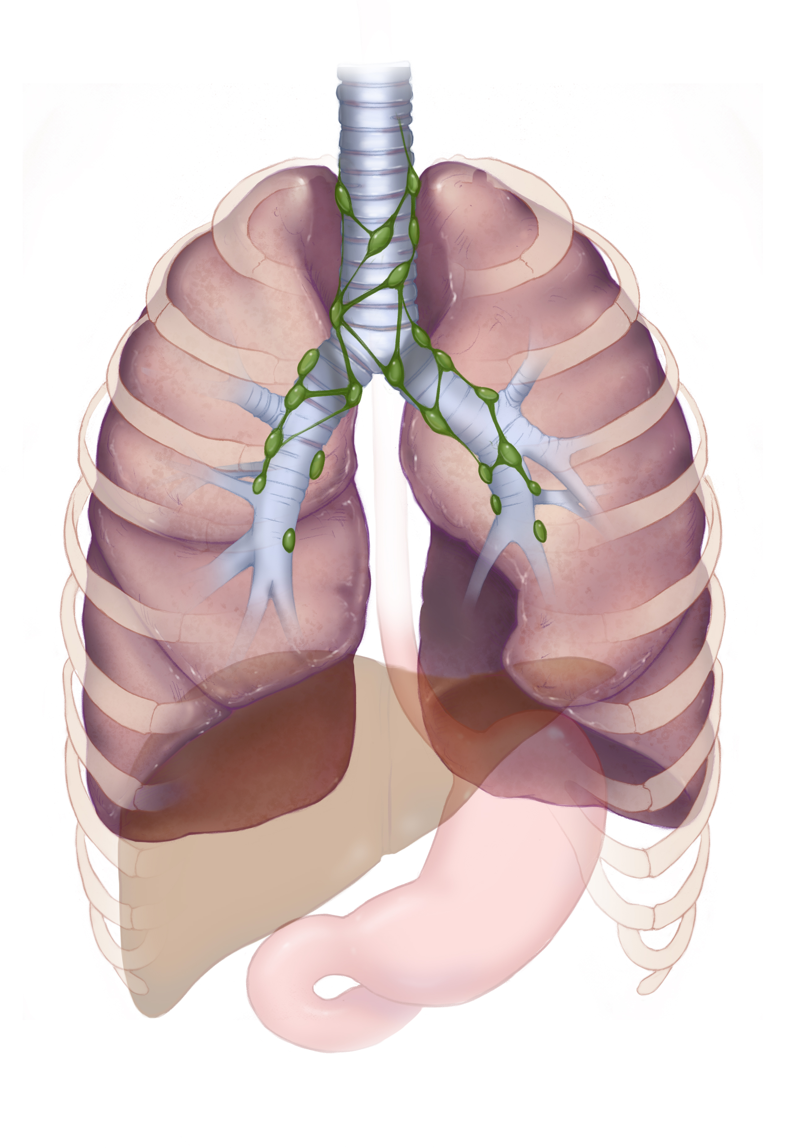
Image-Guided Biopsy of Mediastinal Masses
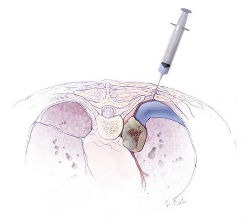
Percutaneous image-guided biopsy of Mediastinal masses: Techniques and Radiologic Pathologic Correlation
Sheila Sheth MD, Syed Ali, MD, Ulrike M Hamper, MD, Frank M. Corl, MS, Elliot K. Fishman MD
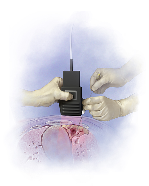
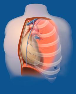
These medical illustrations depict two common image-guided biopsy procedures used to diagnose the nature of mediastinal masses. These illustrations were part of a scientific exhibit discussing the details, techniques, and pitfalls using axial (CT), ultrasound, and endoscopic ultrasound to enable safe access to the lesion, avoidance of vasculature, minimizing risk of pneumothorax, and obtaining adequate lesion samples. These illustrations serve as educational an eye-catching imagery used on the RSNA poster exhibit, as well as a dynamic full-page color image accompanying the radiological journal publication. The primary focus is the education of Radiologists, Oncologists, and Ultrasound Technicians associated with these and other related procedures.
Mediastinum
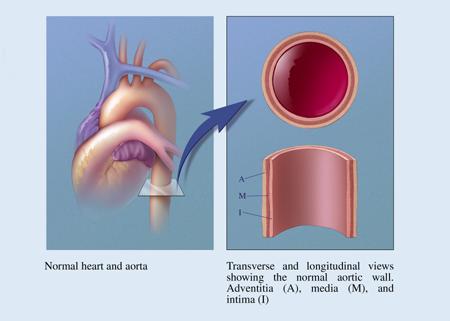
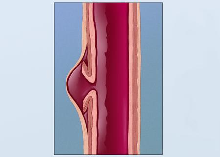
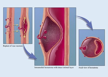
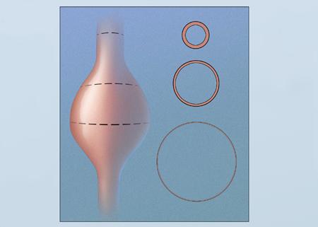
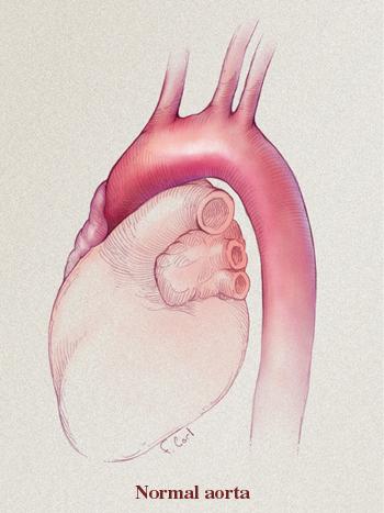
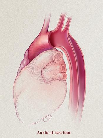
Pathology of the Thoracic Aorta
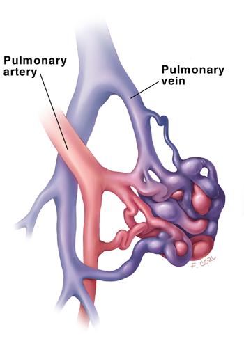
Pulmonary Ateriovenous Malformations (PAVM)
Recurrent Laryngeal Brachial Plexus
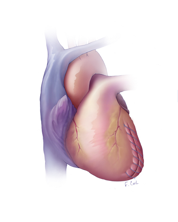
Surgical Ventricular Restoration (SVR)
Surgical Ventricular Restoration: Morphologic and Functional Evaluation Using Multidimensional 16 Detector row CT
Lawler LP, Pannu HK, Conte JV, Corl FM, Fishman EK
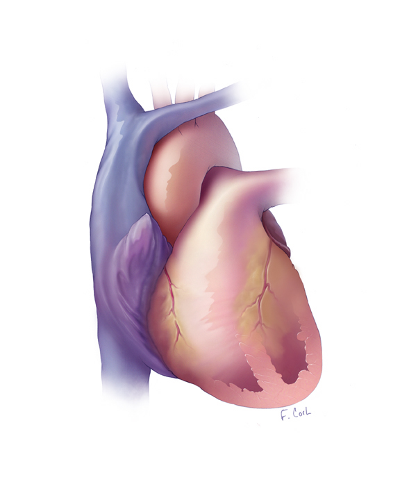
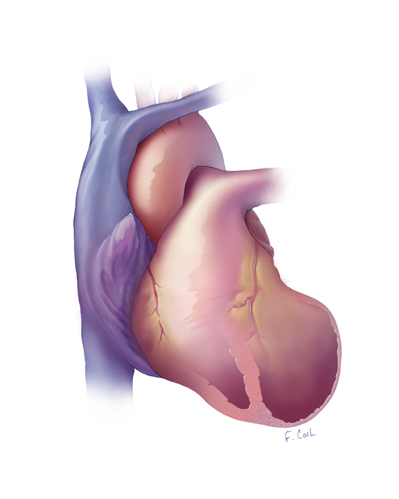
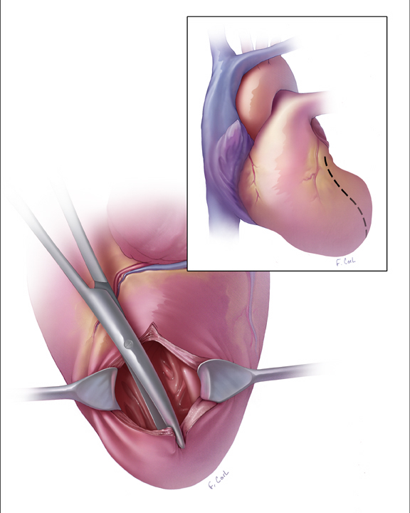
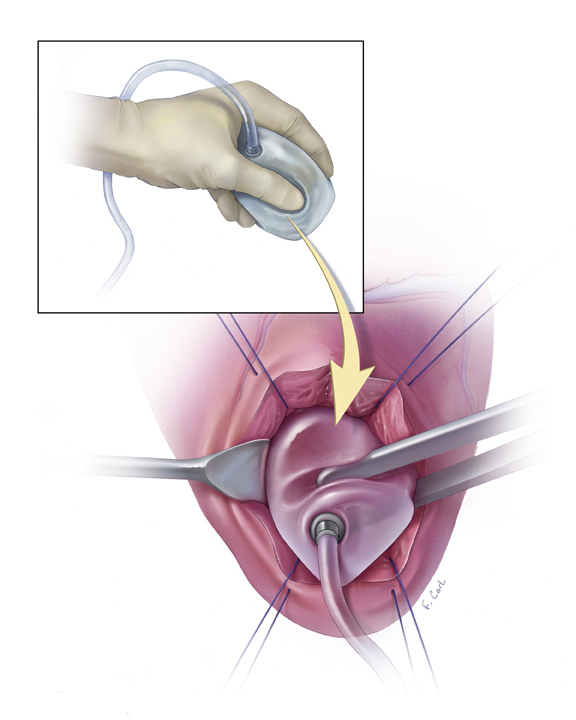
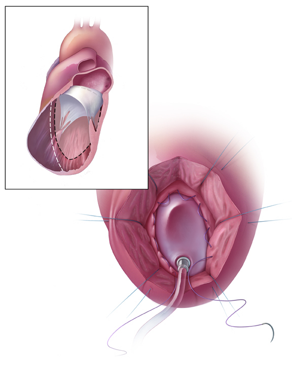
These medical illustrations depict in detail the primary steps of left ventricular restoration, a cutting edge surgical procedure used to improve the lives of patients suffering from congestive heart failure. SVR restores the diseased heart to its normal size and shape, reduces volume in the anterior and septal regions of the left ventricle, and excludes the akinetic and dyskinetic portion of the muscular wall. These illustrations were created for an RSNA scientific poster exhibit and medical journals used to educate radiologists, cardiac surgeons, and other medical professionals associated with this procedure.

