Aortic and Mitral Valve Disease
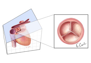
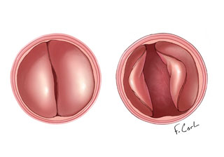
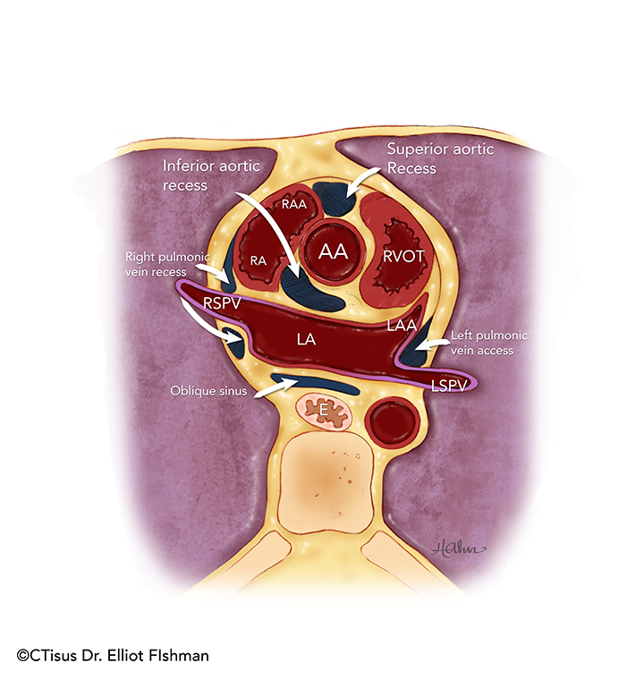
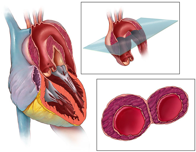
Gated Cardiac Imaging of the Aortic Valve on 64 Slice Multidetector Row Computed Tomography: Preliminary Observations
Pannu HK, Jacobs JE, Shenghan L, Fishman EK
JCAT, 30:3, 443-446, 2006
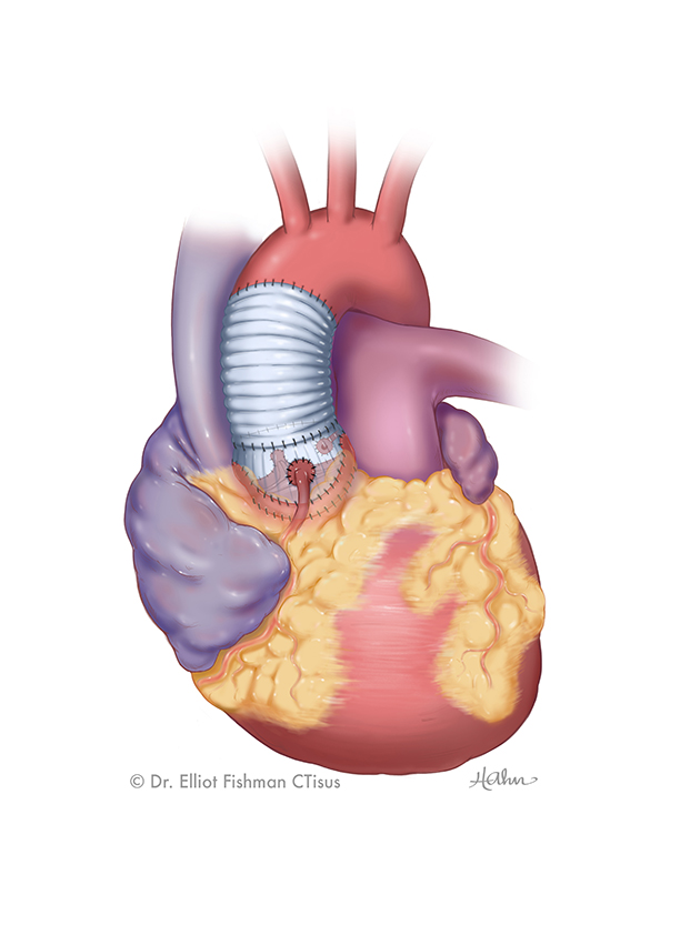
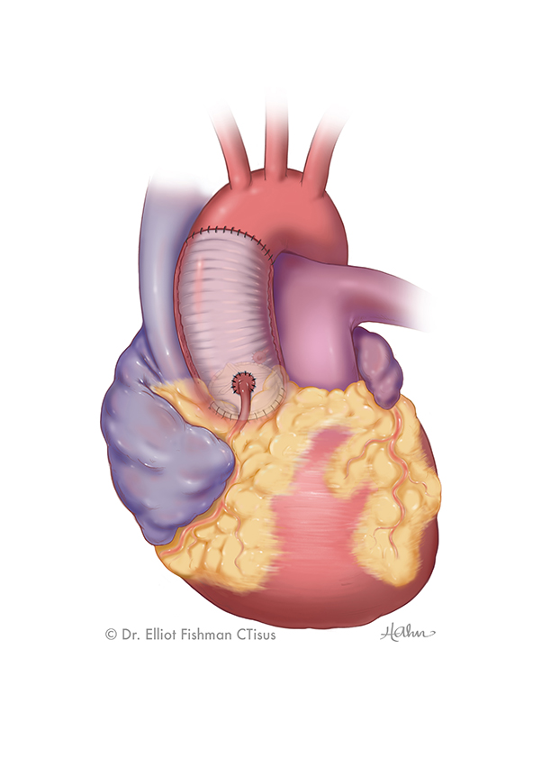
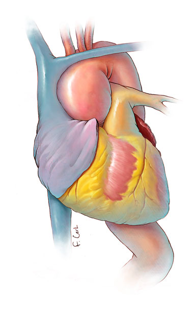
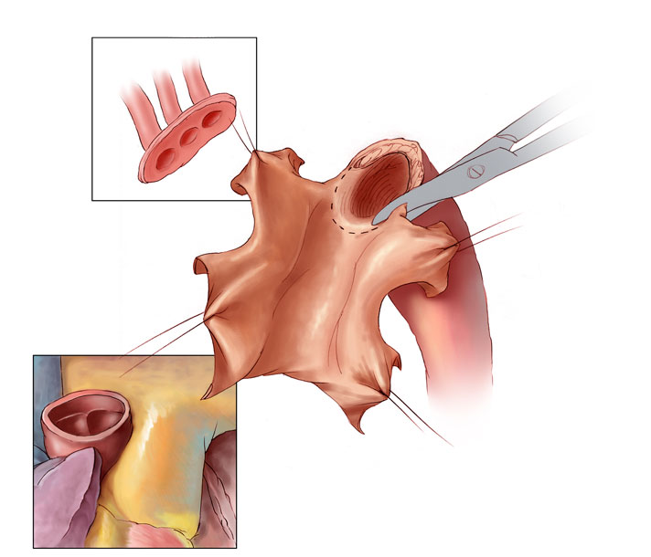
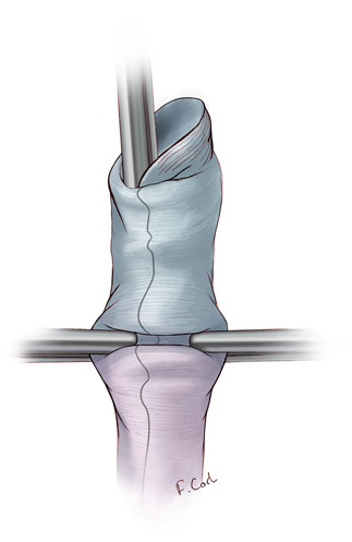
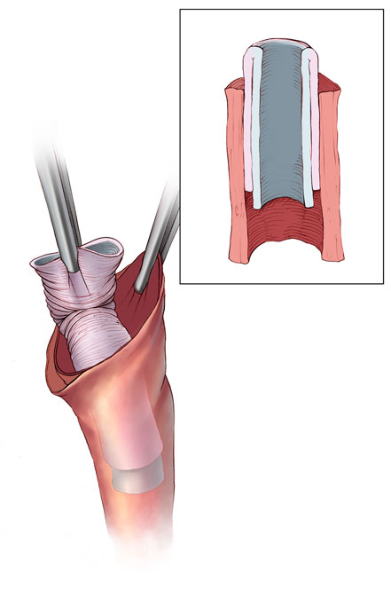
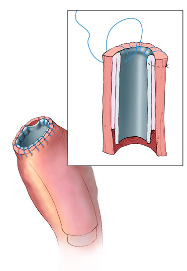
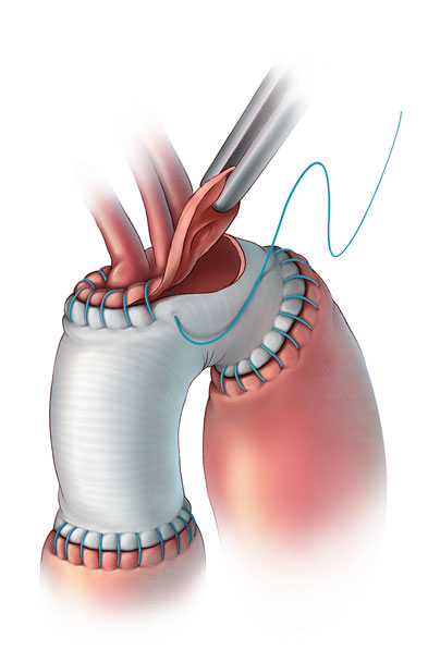
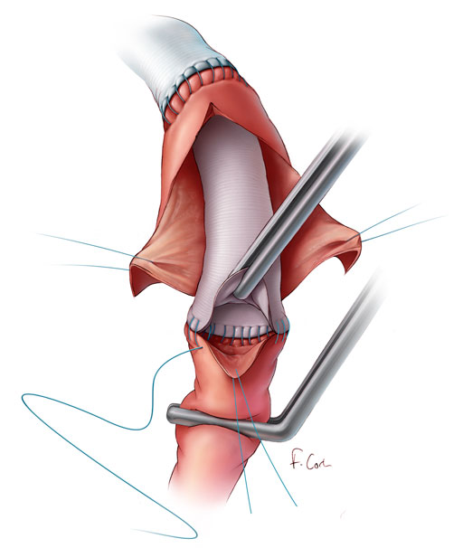
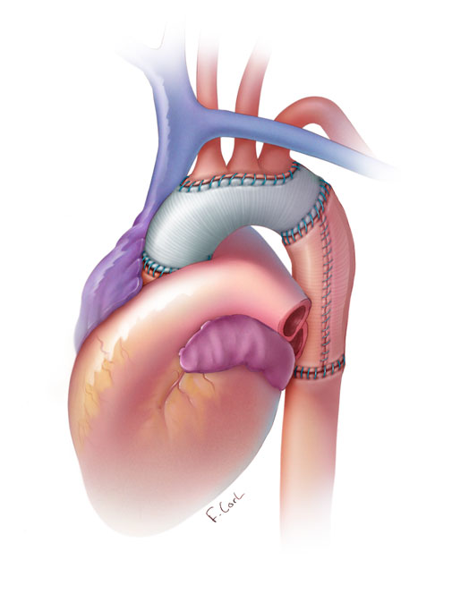
Aortic Root Replacement
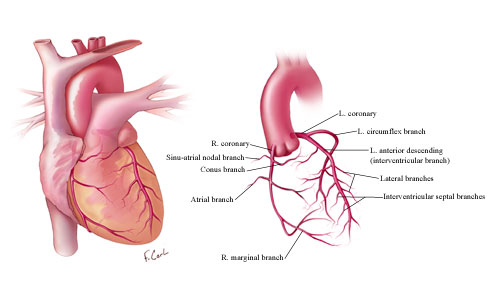
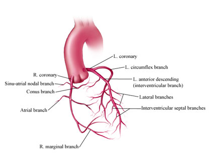
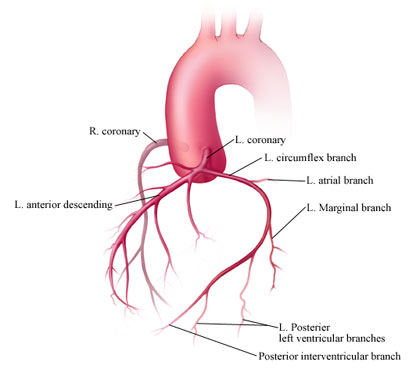
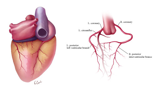
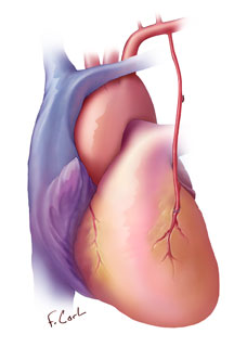
Coronary Artery Anatomy
An Interactive Atlas of Coronary Artery Anatomy and Pathology Based on 64 Slice MDCT, 3D Volume Rendering and Medical Illustration
Frank M. Corl, Harpreet Pannu, Melissa R. Garland, Elliot K. Fishman
These medical illustrations were part of an interactive computer exhibit at RSNA that showed detailed and schematic medical illustrations, axial CT, state of the art 3D MDCT volume rendered images, and animation of normal, aberrant, and pathologic coronary artery anatomy. This exhibit also demonstrated complex anatomy of the coronary arteries in 3 dimensions to better help radiologists that have had limited training with imaging of these vessels.
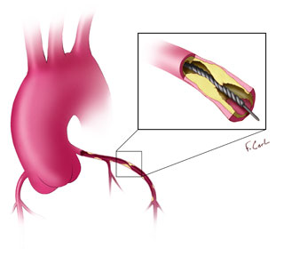
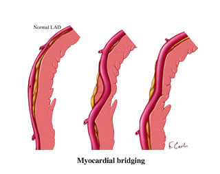
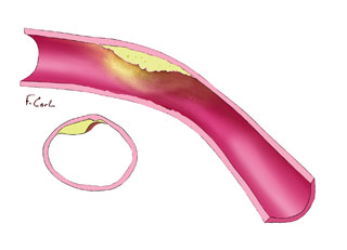
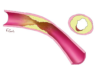
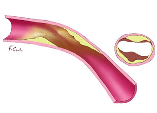
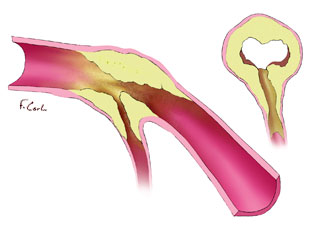
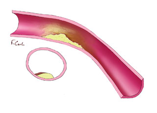
Coronary Artery Pathology
These medical illustrations show very specific types of coronary disease, measurement protocols, and variant anatomy.
MDCT Cardiac CT Protocol
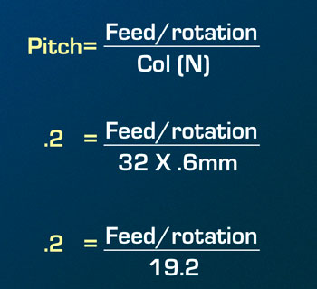
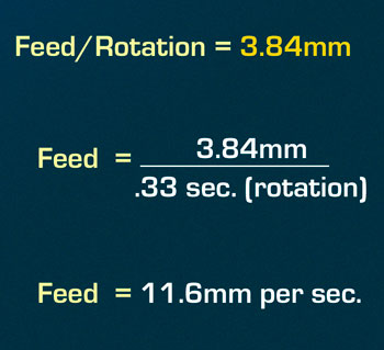
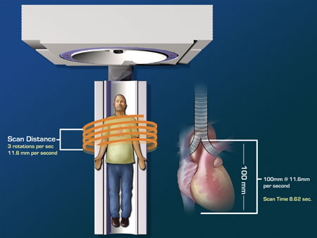
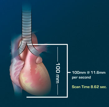
These medical illustrations show a MDCT cardiac CT scanning protocol, including scan area, scan time, pitch, and feed.
Pediatric Congenital Heart Disease
Computerized Tomographic (CT) Imaging of Congenital Heart Disease
Philip J. Spevak MD, Frank Corl MS, Elliot Fishman MD
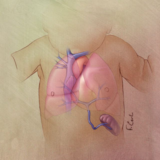
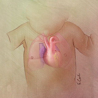
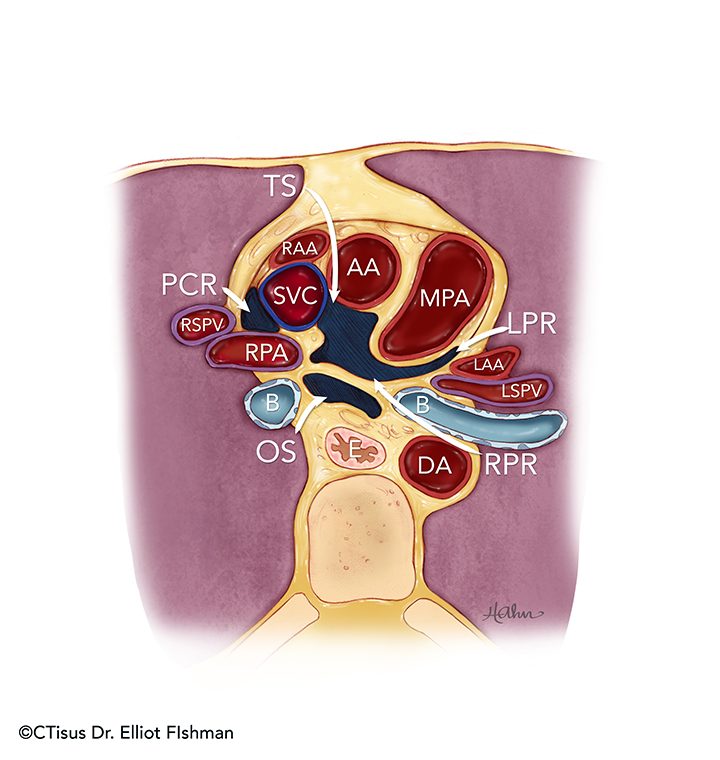
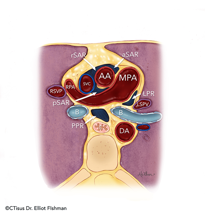
These medical illustrations were part of an RSNA exhibit and journal publication discussing cardiac CT and its role in the imaging of the patient with Congenital Heart Disease.

