- 2
- ,
- 3
- 8
- 1
To Quiz Yourself: Select OFF by clicking the button to hide the diagnosis & additional resources under the case.
Quick Browser: Select ON by clicking the button to hide the additional resources for faster case review.
CASE NUMBER
389
Diagnosis
Pituitary Microadenoma
Note
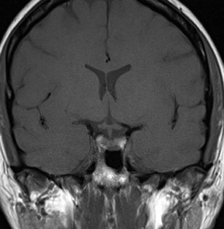
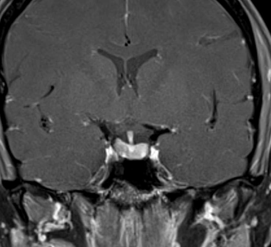
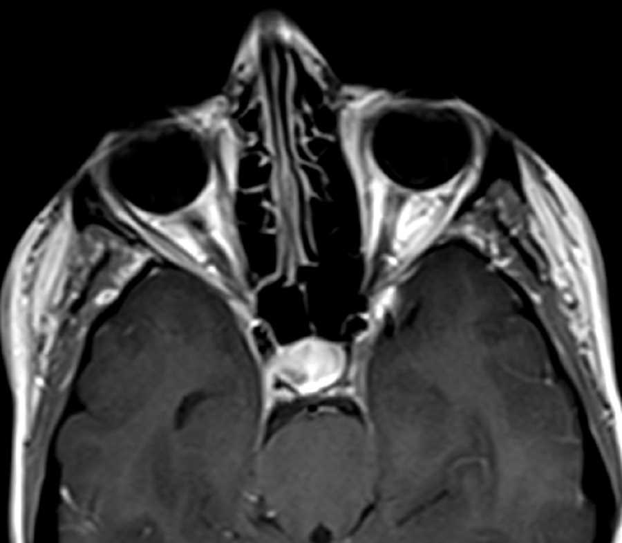
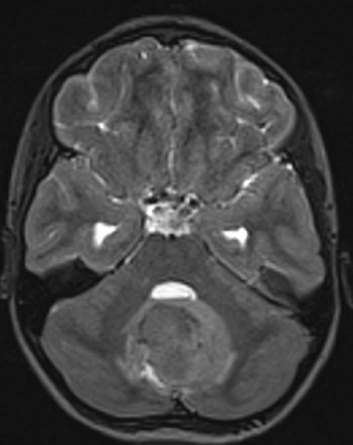
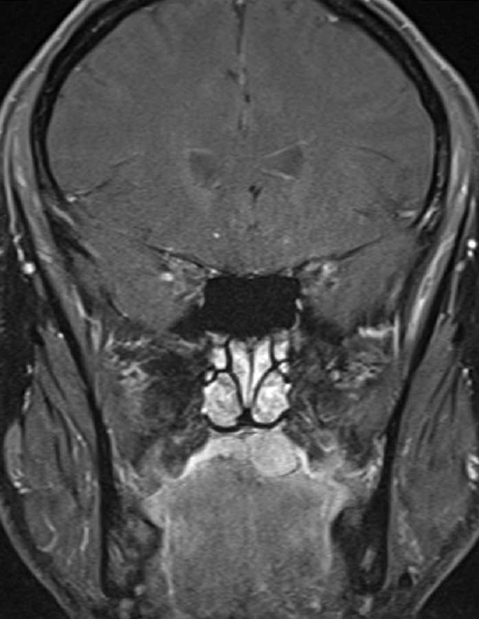
These images show a T1 slightly hypointense, hypoenhancing mass in the posterior right aspect of the pituitary gland, best seen on the sagittal and axial T1 post contrast images. There is no evidence of cavernous sinus invasion. Findings are most compatible with a pituitary microadenoma in this patient with hyperprolactinemia. Differential considerations would include a rathke cleft cyst. Prolactinoma is the most common functional adenoma of the pituitary. In general, microadenomas enhance more slowly than normal pituitary tissue and dynamic post-contrast imaging can be very helpful in identifying subcentimeter lesions.
THIS IS CASE
389
OF
396

