- 2
- ,
- 3
- 8
- 1
To Quiz Yourself: Select OFF by clicking the button to hide the diagnosis & additional resources under the case.
Quick Browser: Select ON by clicking the button to hide the additional resources for faster case review.
CASE NUMBER
390
Diagnosis
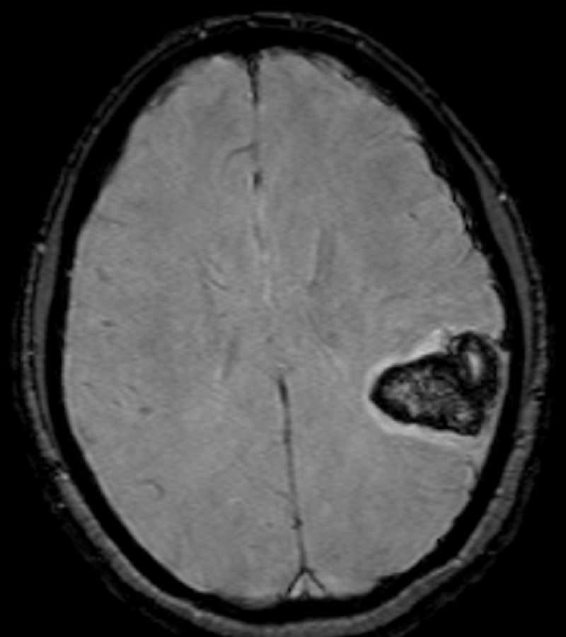
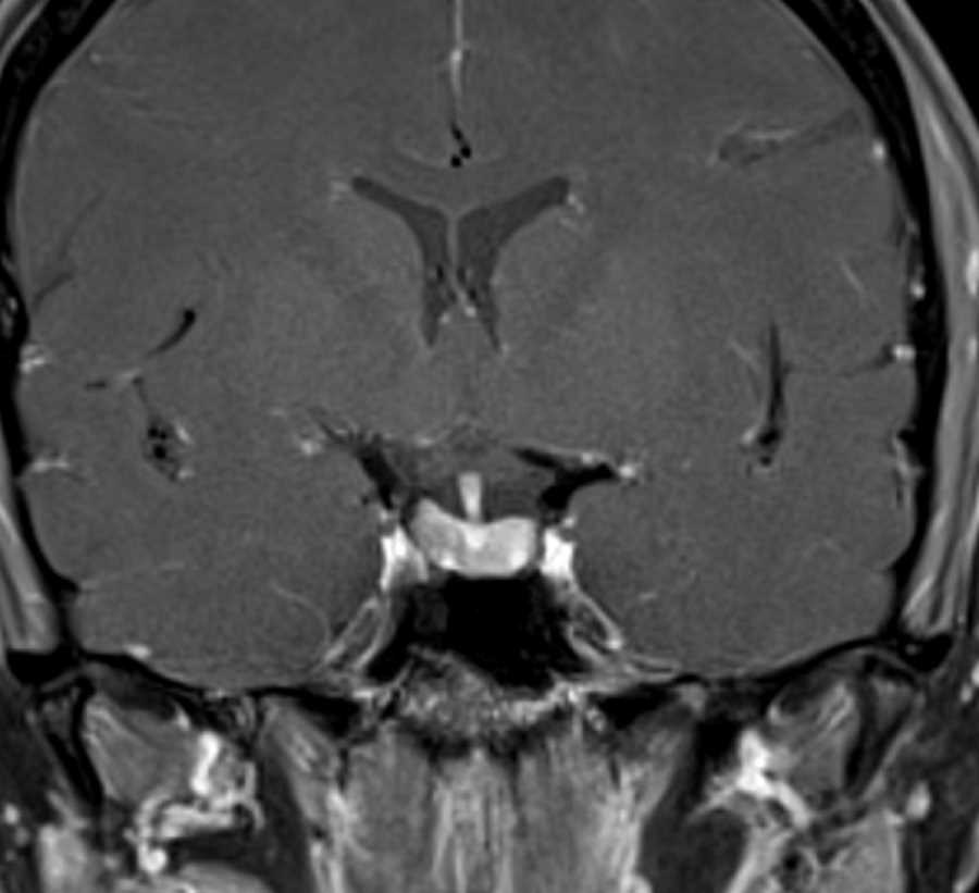
Hemorrhagic Arteriovenous Malformation
Note
These images demonstrate hemorrhage in the left parieto-temporal region best demonstrated on the susceptibility weighted images. Coronal MRV post-contrast images show a small nidus compatible with an arteriovenous malformation. The differential diagnosis on the CT where only hemorrhage was identified included hemorrhagic venous infarction in this young patient. In an elderly patient, the differential for lobar hemorrhage would include amyloid angiopathy. Subsequent catheter angiography showed the nidus to be supplied by two enlarged cortical branches of the superior division of the left middle cerebral artery.
THIS IS CASE
390
OF
396


