- 2
- ,
- 2
- 4
- 6
To Quiz Yourself: Select OFF by clicking the button to hide the diagnosis & additional resources under the case.
Quick Browser: Select ON by clicking the button to hide the additional resources for faster case review.
CASE NUMBER
348


Diagnosis
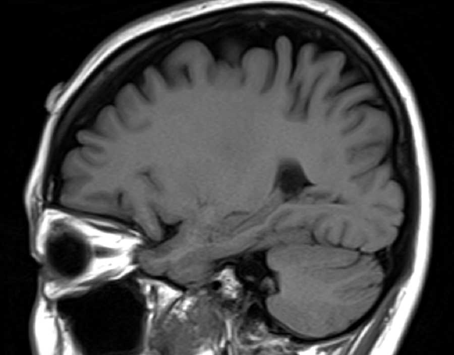
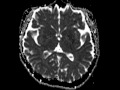
Choroid Plexus Xanthogranuloma
Note
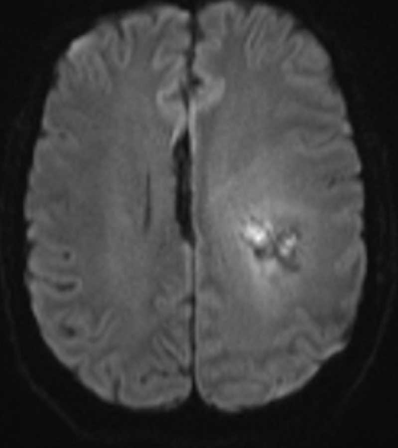
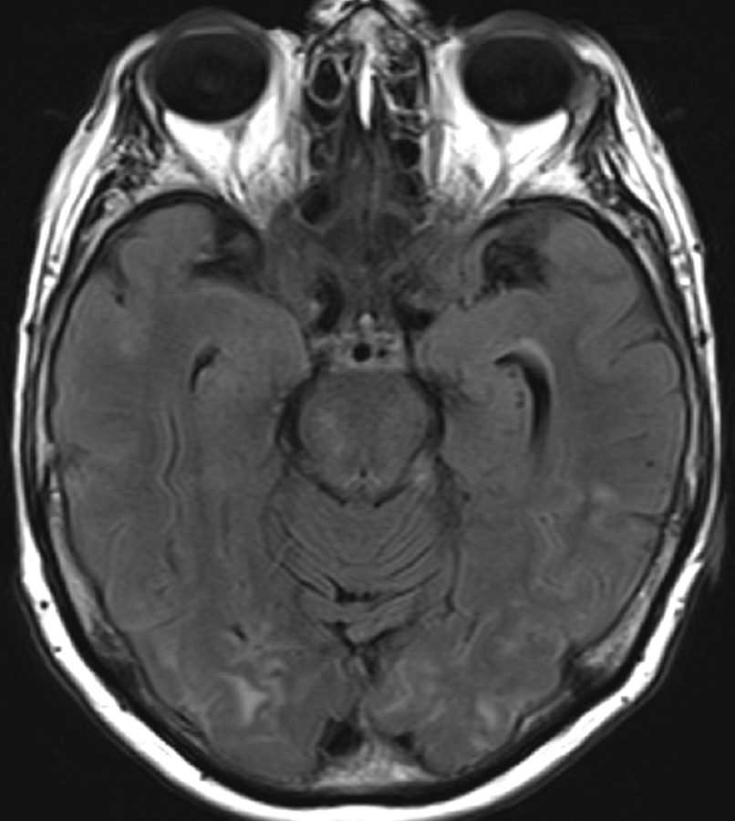
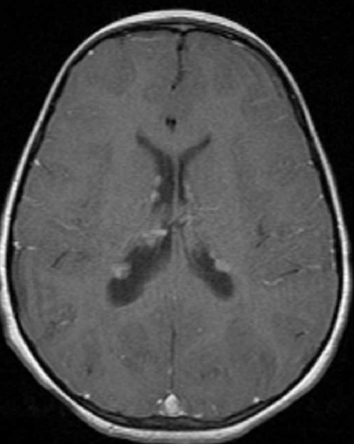
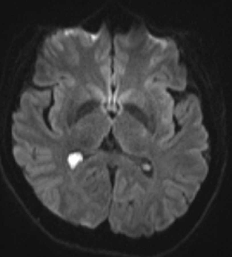
These images show a T2 hyperintense, T2 FLAIR hyperintense, T1 hypointense, non-enhancing mass in the atrium of the right lateral ventricle which is bright on diffusion weighted imaging and has a central area of low ADC value compatible with restricted diffusion. The differential diagnosis may include a parasitic cyst, arachnoid cyst, or choroid plexus cyst or xanthogranuloma. This is a choroid plexus xanthogranuloma which is thought to result from cystic degeneration of the choroid plexus with possible hemorrhage which ultimately fills with cholesterols crystals, calcific deposits, and macrophages resulting in a gelatinous cyst. The restricted diffusion is likely secondary to the high protein content and should not be confused for a neoplasm.
Related videos to the case

THIS IS CASE
348
OF
373