- 2
- ,
- 2
- 4
- 6
To Quiz Yourself: Select OFF by clicking the button to hide the diagnosis & additional resources under the case.
Quick Browser: Select ON by clicking the button to hide the additional resources for faster case review.
CASE NUMBER
347


Diagnosis
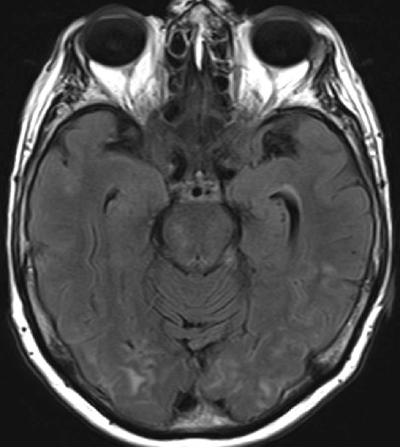
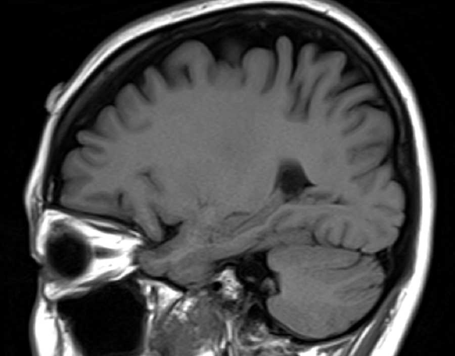
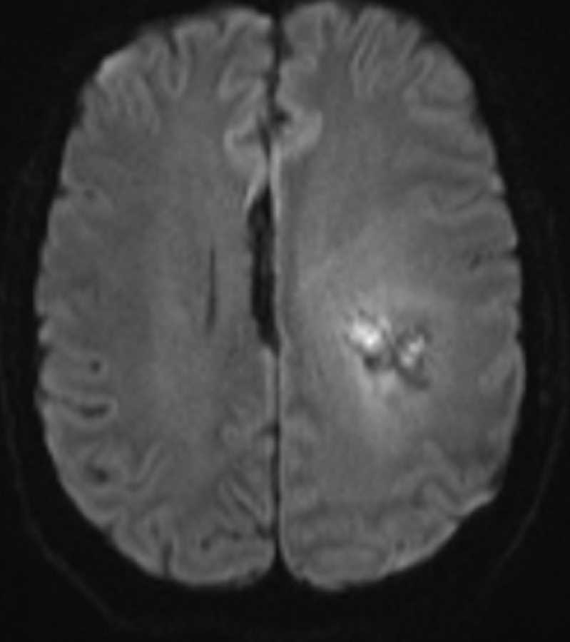
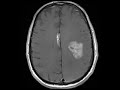
Pericallosal Lipoma
Note
These images show a midline pericallosal T1 hyperintense, T2 FLAIR hyperintense mass which is hypointense on T2 fat saturated images compatible with a lipoma. This finding is incidental as the patient is being followed for the larger, more ominous heterogeneously enhancing mass with perilesional edema in the left cerebral hemisphere compatible with a glioblastoma. The pericallosal location for this lipoma is the most common site for intracranial lipomas and can be seen with anomalies of the corpus callosum.
Related videos to the case

THIS IS CASE
347
OF
373