- 2
- ,
- 2
- 4
- 6
To Quiz Yourself: Select OFF by clicking the button to hide the diagnosis & additional resources under the case.
Quick Browser: Select ON by clicking the button to hide the additional resources for faster case review.
CASE NUMBER
346


Diagnosis
Amyloid Angiopathy
Note
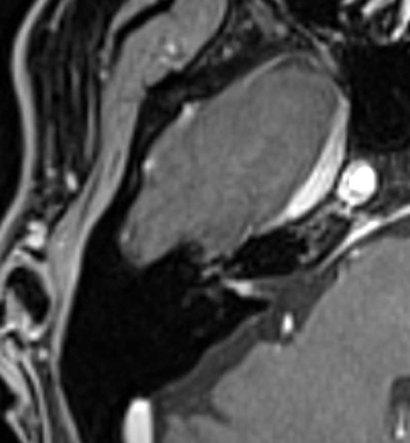
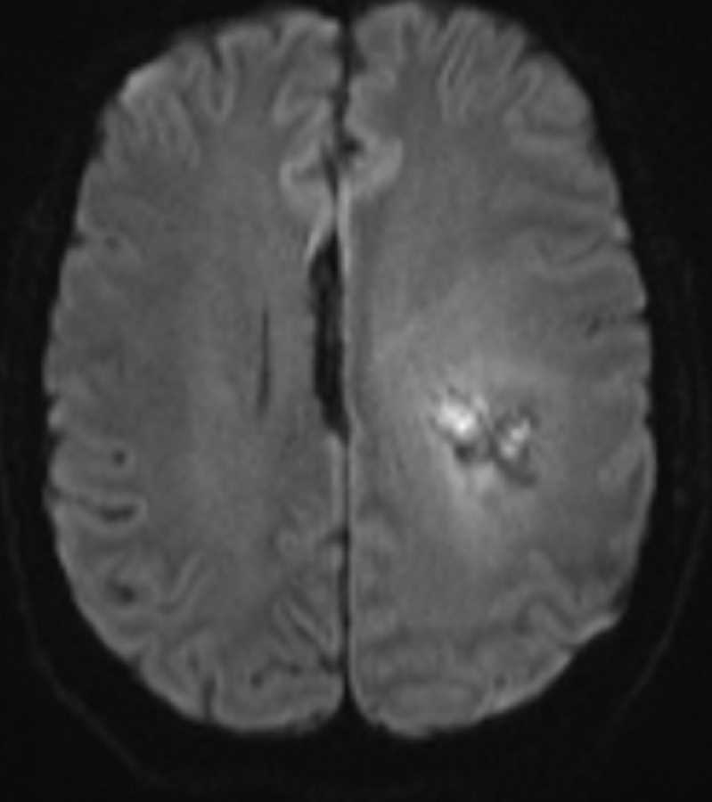
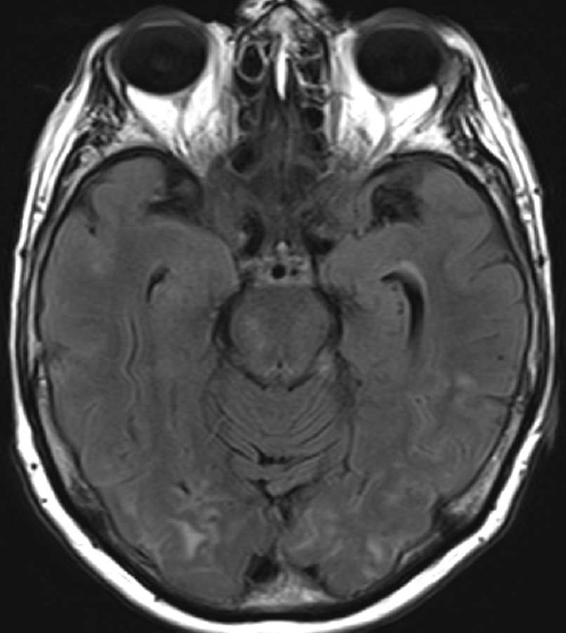
These images demonstrate innumerable foci of increased susceptibility throughout the cerebral hemispheres and cerebellum, relatively sparing the anterior frontal lobes and brainstem. These foci are associated with areas of T2 FLAIR hyperintensity and are compatible with remote microhemorrhages. The differential diagnosis includes multiple cavernomas, traumatic diffuse axonal injury, amyloid angiopathy, posterior reversible encephalopathy, and hypertensive microhemorrhages. The best diagnosis in this elderly patient is amyloid angiopathy. The parietal and occipital lobes are most commonly involved as well as the cortical and subcortical location. Lobar hemorrhage not seen here is another characteristic feature of cerebral amyloid disease.
Related videos to the case

THIS IS CASE
346
OF
373