- 2
- ,
- 2
- 4
- 6
To Quiz Yourself: Select OFF by clicking the button to hide the diagnosis & additional resources under the case.
Quick Browser: Select ON by clicking the button to hide the additional resources for faster case review.
CASE NUMBER
345


Diagnosis
Epidermoid
Note
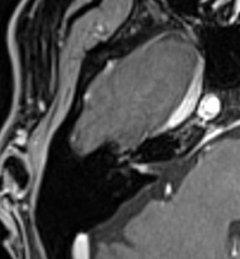
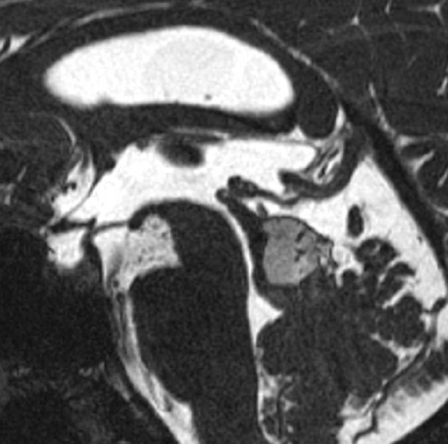
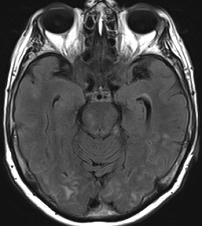
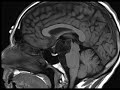
These images demonstrate a lobulated T2 hyperintense, T1 hypointense, T2 FLAIR hyperintense, non-enhancing mass centered in the suprasellar and interpeduncular cisterns. The mass demonstrates restricted diffusion and is compatible with an epidermoid cyst. Most epidermoids are intradural and commonly involve the basilar cisterns as seen here. They tend to follow the signal of CSF, but do not completely suppress on FLAIR. Diffusion restriction is the defining imaging feature. Treatment is with surgical resection.
Related videos to the case

THIS IS CASE
345
OF
373