- 2
- ,
- 2
- 4
- 6
To Quiz Yourself: Select OFF by clicking the button to hide the diagnosis & additional resources under the case.
Quick Browser: Select ON by clicking the button to hide the additional resources for faster case review.
CASE NUMBER
340


Diagnosis
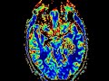
Intraventricular Metastasis
Note
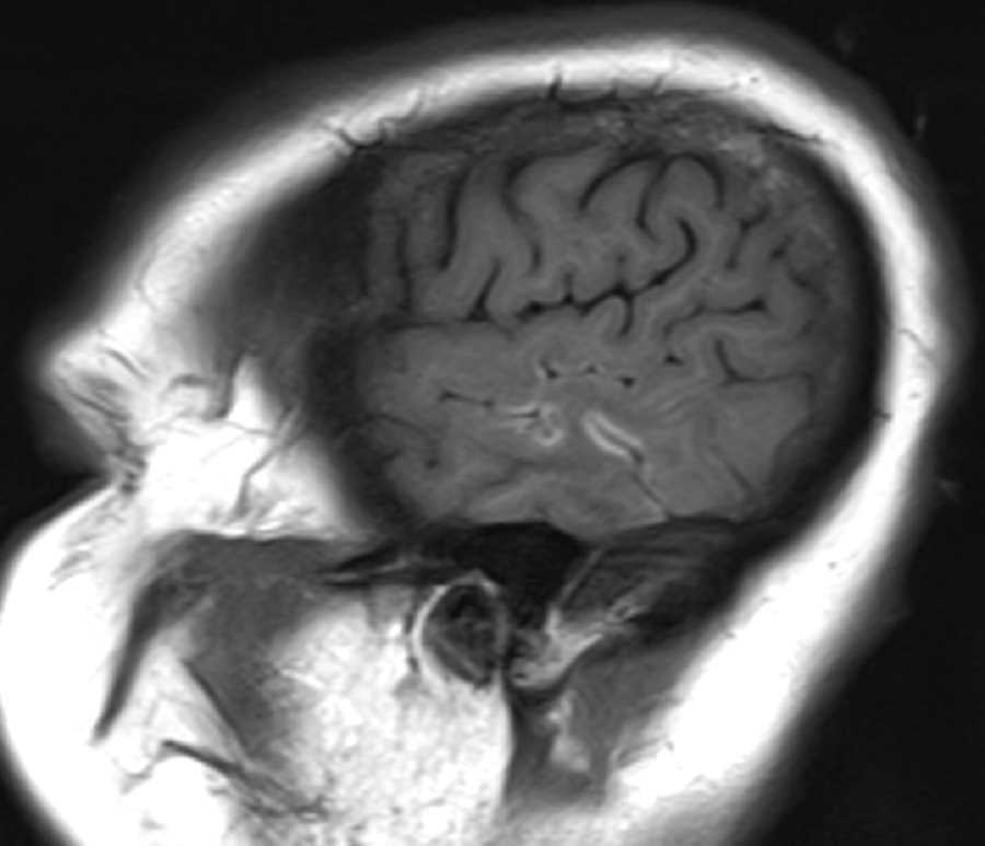
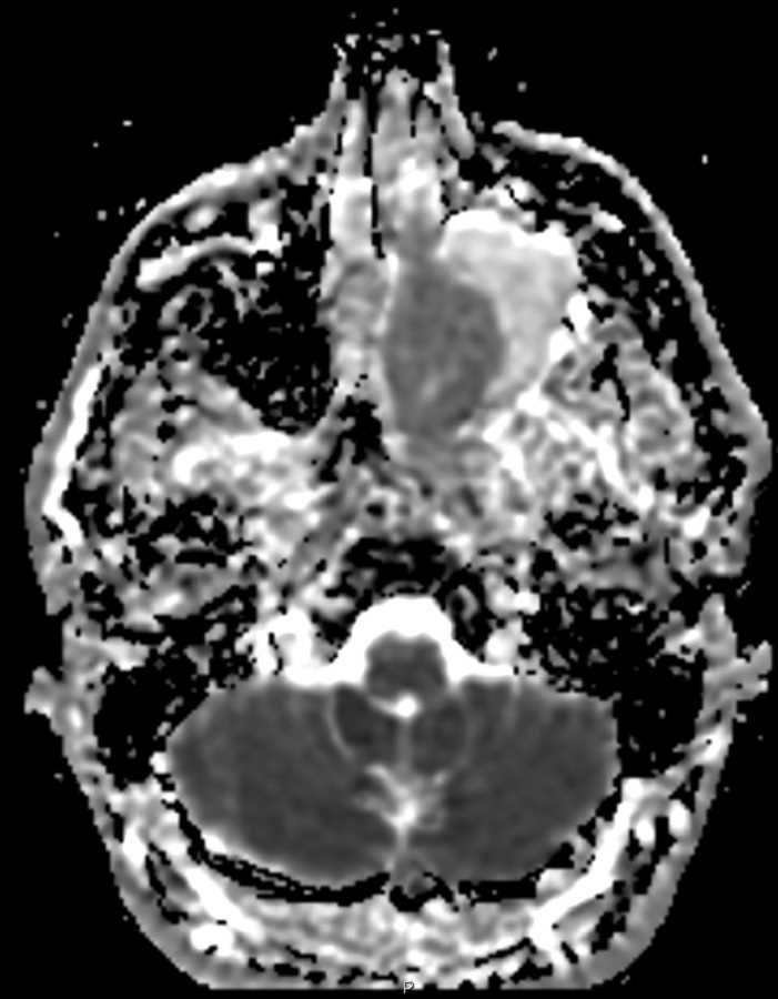
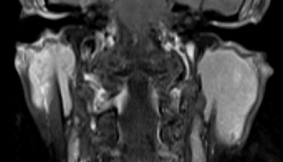
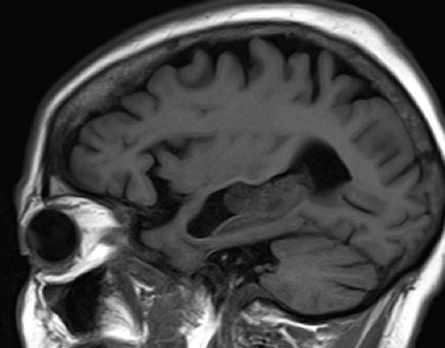
Centered in the mid temporal horn of the left lateral ventricle, there is a multilobulated T1 isointense, T2 hypointense, heterogeneously enhancing mass in contiguity with the choroid plexus. The marked T2 hypointensity is indicative of hemorrhage. Perfusion weighted imaging shows increased perfusion. The distal temporal horn is asymmetrically dilated compatible with entrapment of CSF by the mass. There is adjacent T2 FLAIR hyperintensity in the left temporal lobe compatible with vasogenic edema. The differential for this lesion includes metastasis, choroid plexus tumor, neurocytoma, or ependymoma. This patient had a history of lung cancer, so metastasis is the best diagnosis. Renal cell carcinoma is another tumor well known to metastasize to the choroid plexus.
Related videos to the case

THIS IS CASE
340
OF
373