- 2
- ,
- 2
- 4
- 6
To Quiz Yourself: Select OFF by clicking the button to hide the diagnosis & additional resources under the case.
Quick Browser: Select ON by clicking the button to hide the additional resources for faster case review.
CASE NUMBER
339


Diagnosis
Neurosarcoidosis
Note
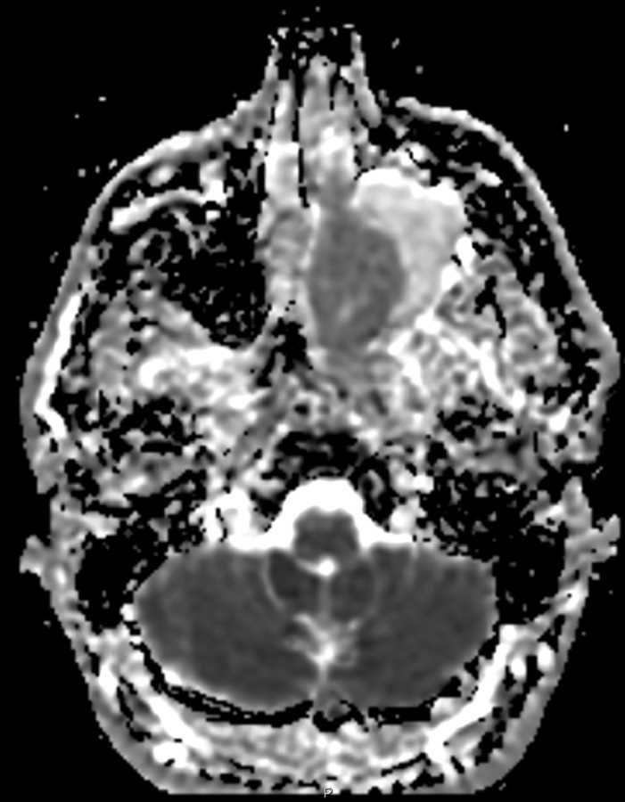
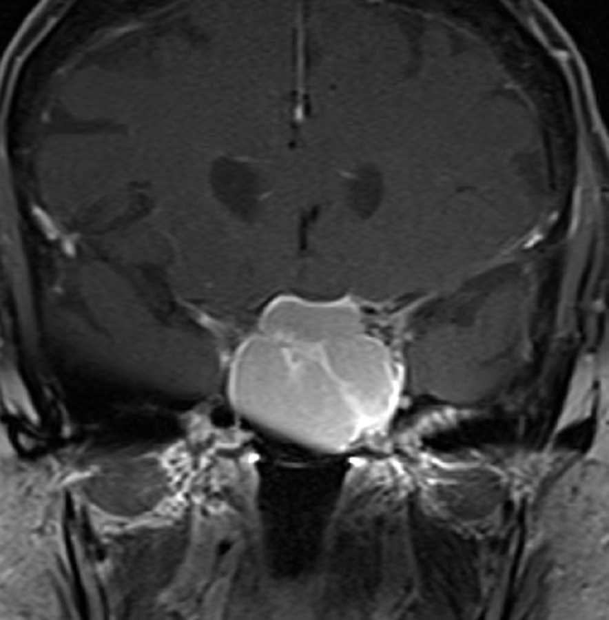
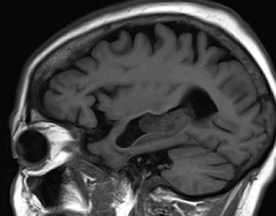
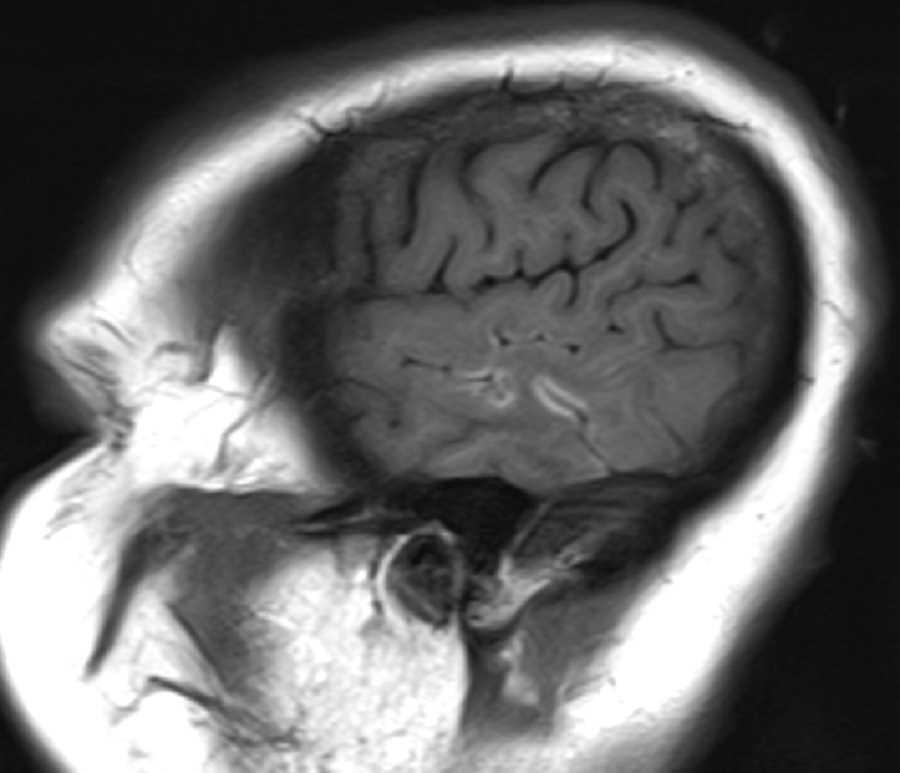
In the left temporal lobe, there is mildly expansile T2 FLAIR hyperintensity, cortical T1 hyperintensity, and nodular leptomeningeal enhancement with few tiny foci of restricted diffusion. Findings are compatible with active inflammation with differential considerations including leptomeningeal metastasis, meningoencephalitis, primary CNS vasculitis with subacute infarction, and granulomatous infection. This is a case of neurosarcoidosis with associated vasculitis resulting in predominantly vasogenic edema with suspected cortical laminar necrosis and few foci of cytotoxic edema. Neurosarcoidosis may have a variety of CNS manifestations. Most commonly it infiltrates the dura and pia of the basilar cisterns and may involve the pituitary stalk, hypothalamus, and brainstem. Involvement of the cerebral hemispheres and the brain parenchyma as seen in this case is less common. Patients usually present with cranial nerve deficits.Treatment consists of managing symptoms with steroids with 50% showing signs of progression despite treatment.
Related videos to the case


THIS IS CASE
339
OF
373