- 2
- ,
- 3
- 8
- 1
To Quiz Yourself: Select OFF by clicking the button to hide the diagnosis & additional resources under the case.
Quick Browser: Select ON by clicking the button to hide the additional resources for faster case review.
CASE NUMBER
358


Diagnosis
Watershed Infarcts
Note
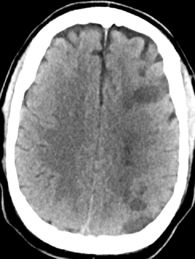
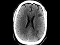
This patient had a history of PEA (pulseless electrical activity) arrest and acute right-sided neurological deficits. The CT shows multifocal areas of wedge-shaped hypoattenuation involving the cortex and subcortical white matter of the left frontal and parietal lobes at the expected junction of the major arterial territories. In addition, there is hypoattenuation of the left centrum semiovale at the expected border of the perforating arteries. Findings are compatible with watershed or hypotensive infarctions which fits clinically with this patients history of cardiac arrest. Given the unilaterality of the findings, the patient may have a stenosis of the left carotid predisposing the left cerebral hemisphere to the ischemic insult following the drop in blood pressure. Differential considerations include embolic infarcts.
Related videos to the case

THIS IS CASE
358
OF
396