- 2
- ,
- 2
- 5
- 2
To Quiz Yourself: Select OFF by clicking the button to hide the diagnosis & additional resources under the case.
Quick Browser: Select ON by clicking the button to hide the additional resources for faster case review.
CASE NUMBER
313


Diagnosis
Periventricular Leukomalacia
Note
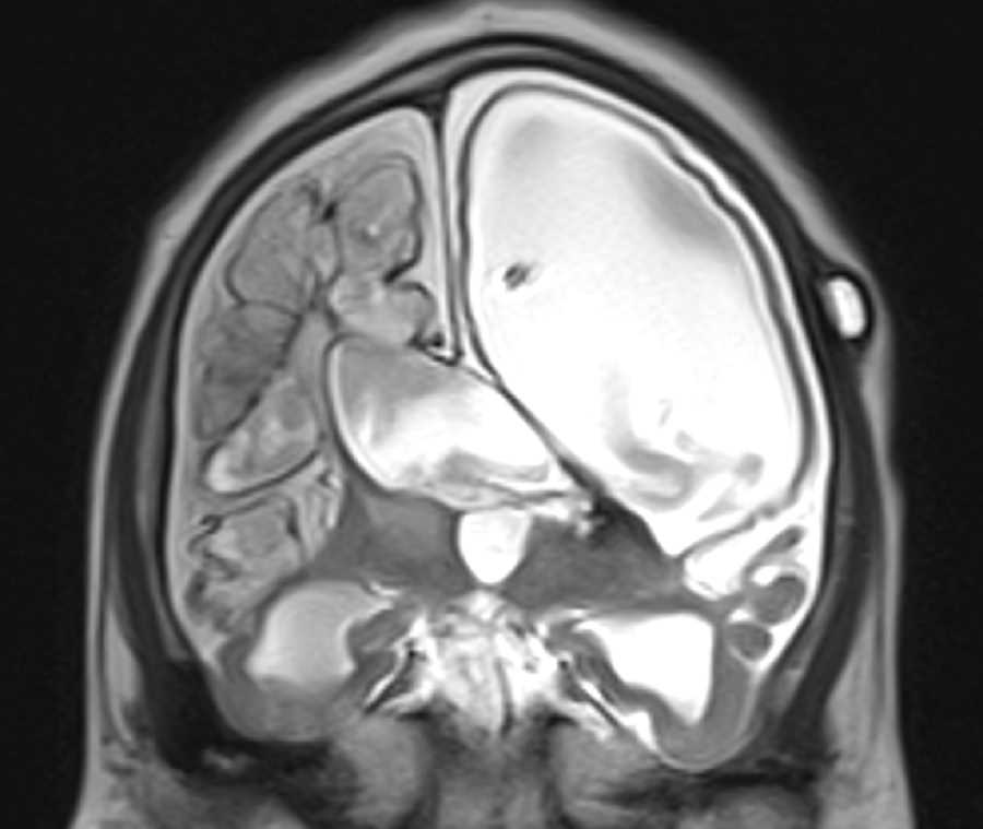
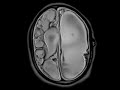
This study was performed as part of an ultrafast protocol for shunted hydrocephalus which consist of multiplanar T2 haste images are obtained in quick fashion to assess changes in ventricular size. These images demonstrate sequelae of severe hypoxic ischemic injury in the neonatal period which resulted in severe periventricular white matter volume loss with ex vacuo enlargement of the lateral ventricles predominantly on the left. There is gray matter volume loss as well. Part of the hemispheric cortex is preserved as a T2 hypointense residual tissue layer. There is a left-sided VP shunt with the tip located within the left lateral ventricle. In addition to the cerebral atrophy, the brainstem and cerebellum demonstrates volume loss with dilatation of the fourth ventricle and widening of the foramina of Luschka, bilaterally. Overall the patient is microcephalic.
Related videos to the case

THIS IS CASE
313
OF
374