- 2
- ,
- 2
- 4
- 6
To Quiz Yourself: Select OFF by clicking the button to hide the diagnosis & additional resources under the case.
Quick Browser: Select ON by clicking the button to hide the additional resources for faster case review.
CASE NUMBER
310


Diagnosis
Meningioma
Note
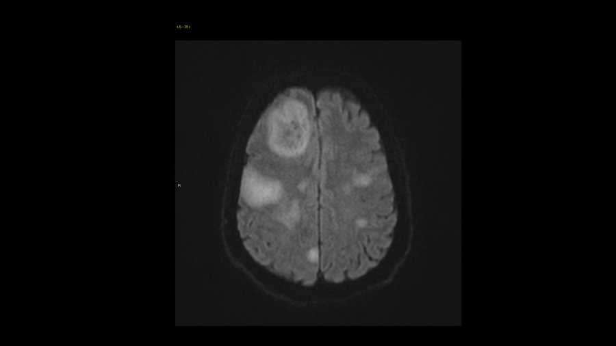
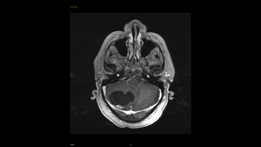
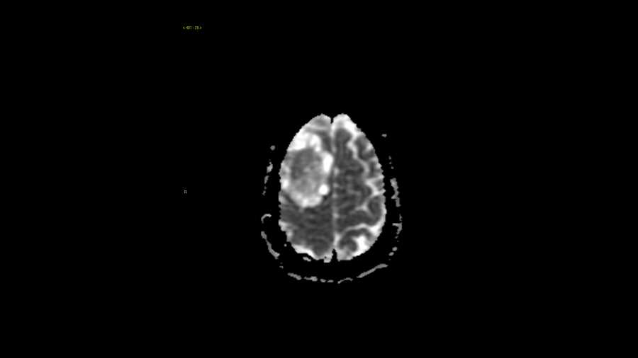
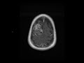
These images show a large T2 hyperintense, multi-septated, cystic, extra-axial mass along the right frontal convexity with significant mass effect on the subjacent brain, but no evidence of edema. This indicates a slow growing process and is compatible with a meningioma. Of importance from a surgical standpoint, there is no evidence of superior sagittal sinus infiltration. Meningiomas can be cystic which may represent areas of degeneration or necrosis. The differential in this case includes a hemangiopericytoma, but these are usually locally aggressive, features which are not seen in this case.
Related videos to the case

THIS IS CASE
310
OF
373