- 2
- ,
- 2
- 5
- 2
To Quiz Yourself: Select OFF by clicking the button to hide the diagnosis & additional resources under the case.
Quick Browser: Select ON by clicking the button to hide the additional resources for faster case review.
CASE NUMBER
312


Diagnosis
Acute Right MCA Infarct
Note
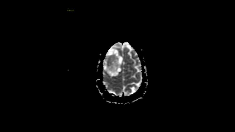
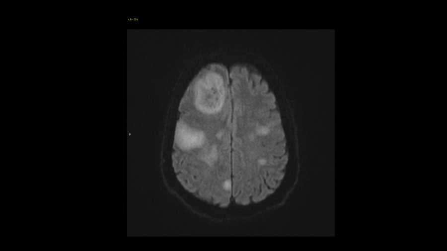
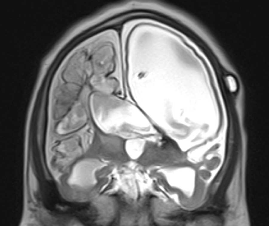
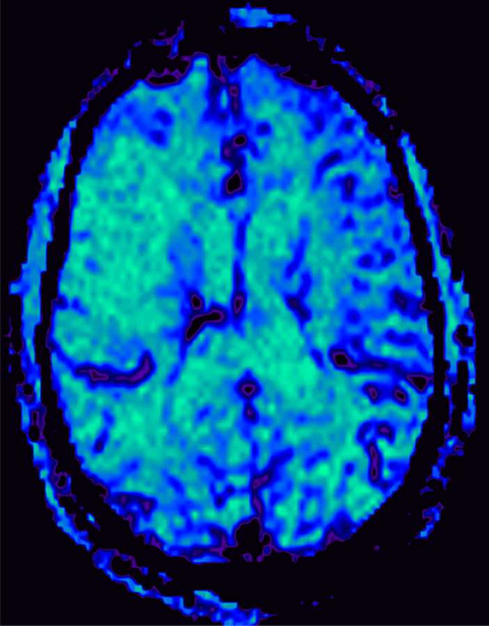
These images show hyperattenuation of the right middle cerebral artery at the M1 and M2 segment junction on CT which is known as the hyperdense MCA sign and signifies clot. There was no discernable loss of gray white matter differentiation on CT at this point and the patient went on to get MRI and MRA of the brain for further characterization of the extent of the acute infarct. MRI showed restricted diffusion throughout the right MCA territory with matching perfusion abnormality and clot in the MCA as evidenced by lack of flow related enhancement on 3D time of flight imaging and blooming artifact on susceptibility weighted imaging. The patient had an old infarct involving the left periatrial white matter with hemosiderin deposition which was also noted on the CT.
Related videos to the case


THIS IS CASE
312
OF
374