- 2
- ,
- 2
- 4
- 6
To Quiz Yourself: Select OFF by clicking the button to hide the diagnosis & additional resources under the case.
Quick Browser: Select ON by clicking the button to hide the additional resources for faster case review.
CASE NUMBER
279


Diagnosis
Cortical Dysplasia
Note
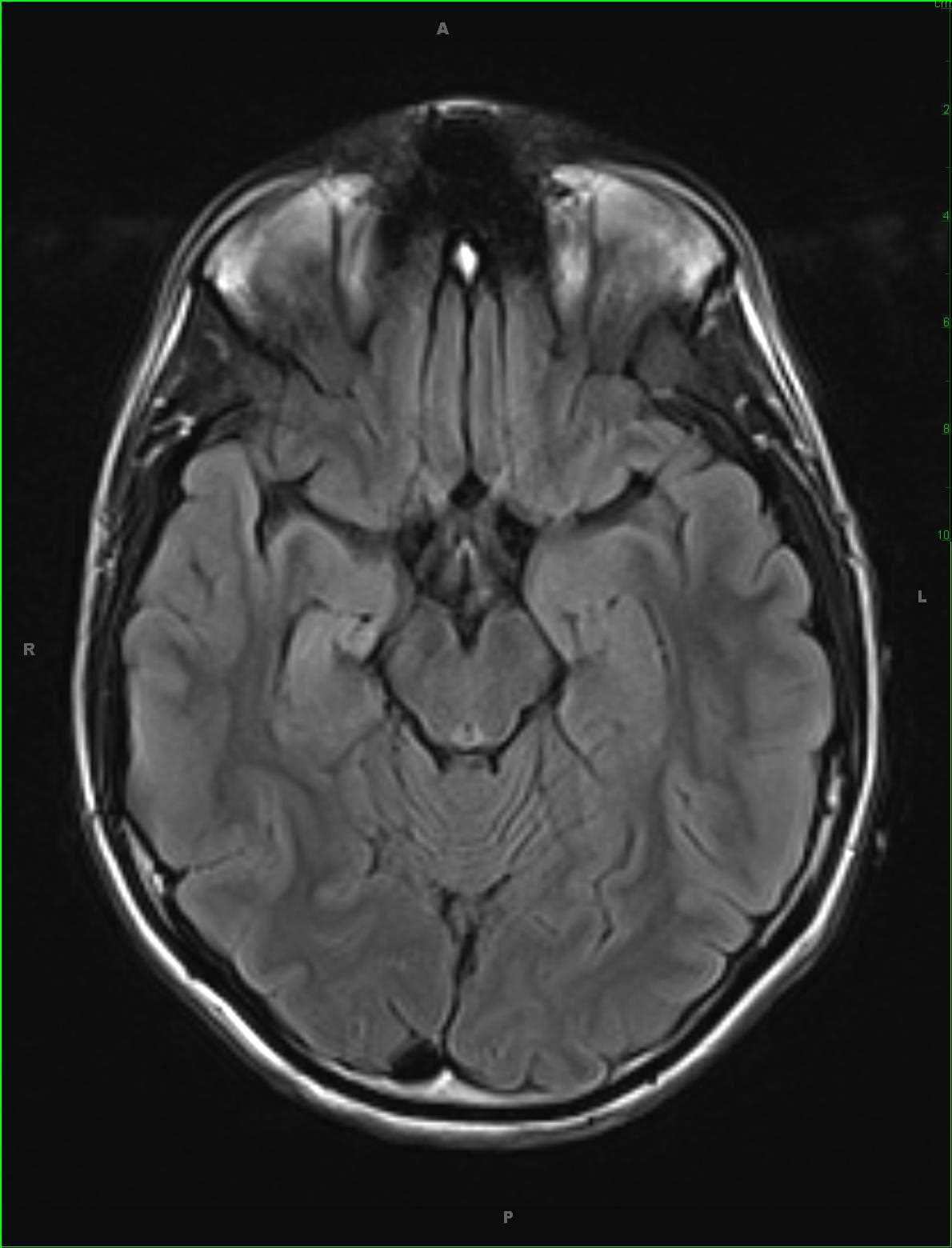
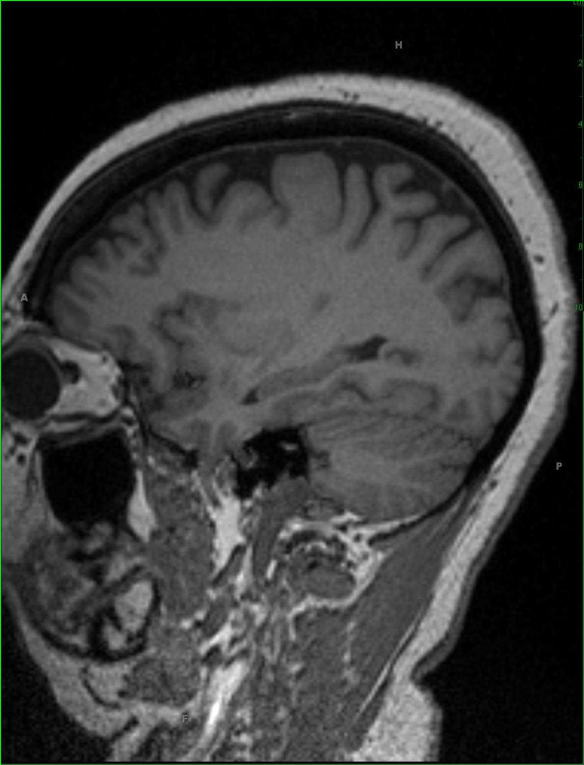
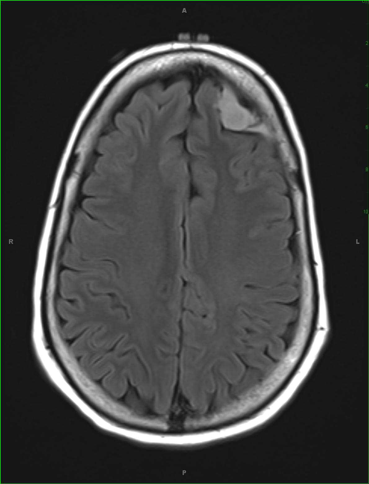
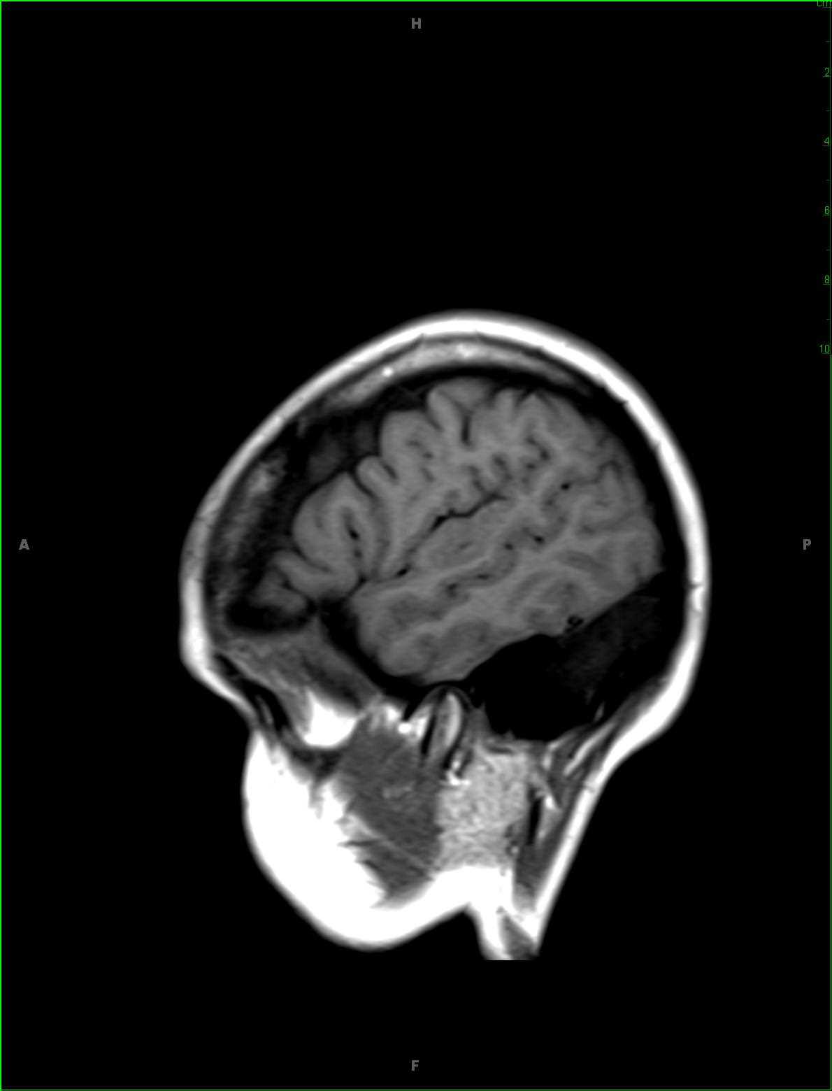
Long-standing history of right temporal lobe epilepsy localizing to the superior temporal gyral region on EEG. There is a disorganized appearance of the cortical gray matter along Heschls gyrus on the right. There is thickening of the cortical gray matter which is indistinct on the more lateral sagittal T1-weighted image. A large confluent region of T2/FLAIR hyperintense signal was identified within the superior right temporal gyrus without diffusion restriction or loss of signal on the susceptibility weighted images. No suspicious contrast enhancement is identified. A differential of cortical dysplasia, dysembryoplastic neuroectodermal tumor, ganglioglioma and low-grade astrocytoma was given. On biopsy, the region of signal abnormality was found to reflect a region of focal cortical dysplasia type Ia. Focal cortical dysplasia (FCD) can be divided into two categories, FCD type I (non-Taylor dysplasia) and FCD type II (Taylor dysplasia). FCD type Ia results from dyslamination and mild malformation of cortical development. FCD type Ib results from isolated architectural abnormalities and cytoarchitectural dysplasia. FCD type II can further be subdivided into type IIa, no balloon cells, and type IIb, balloon cells present.
Related videos to the case