- 2
- ,
- 2
- 4
- 6
To Quiz Yourself: Select OFF by clicking the button to hide the diagnosis & additional resources under the case.
Quick Browser: Select ON by clicking the button to hide the additional resources for faster case review.
CASE NUMBER
278


Diagnosis
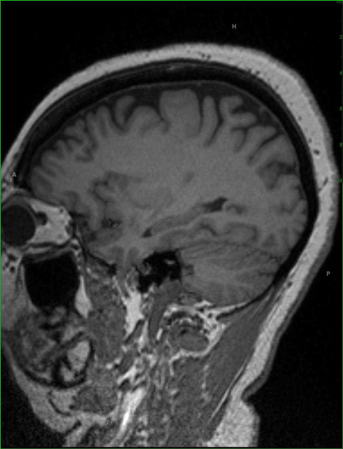
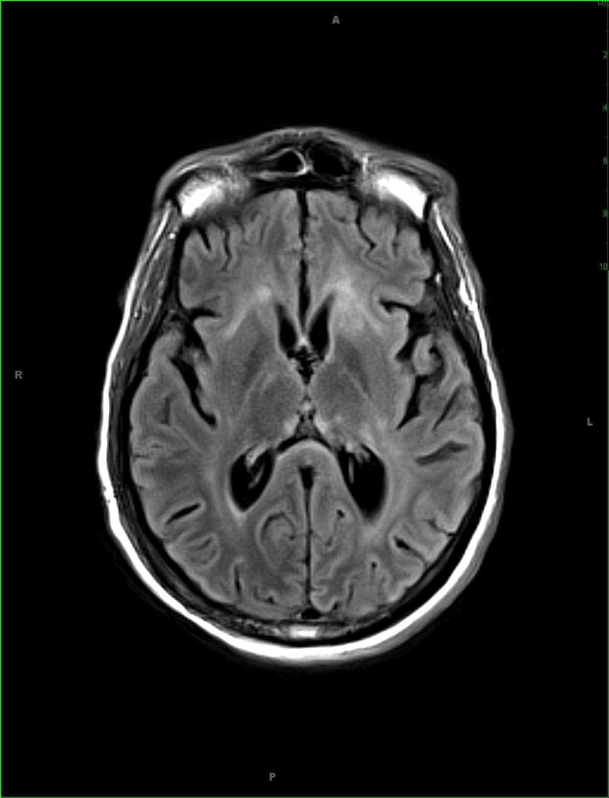
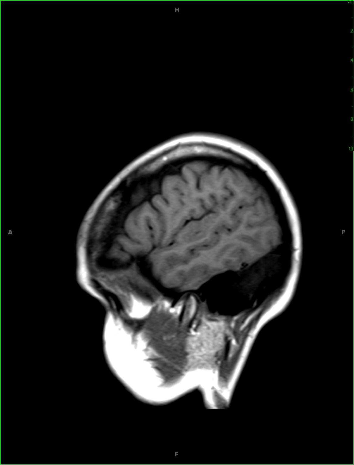
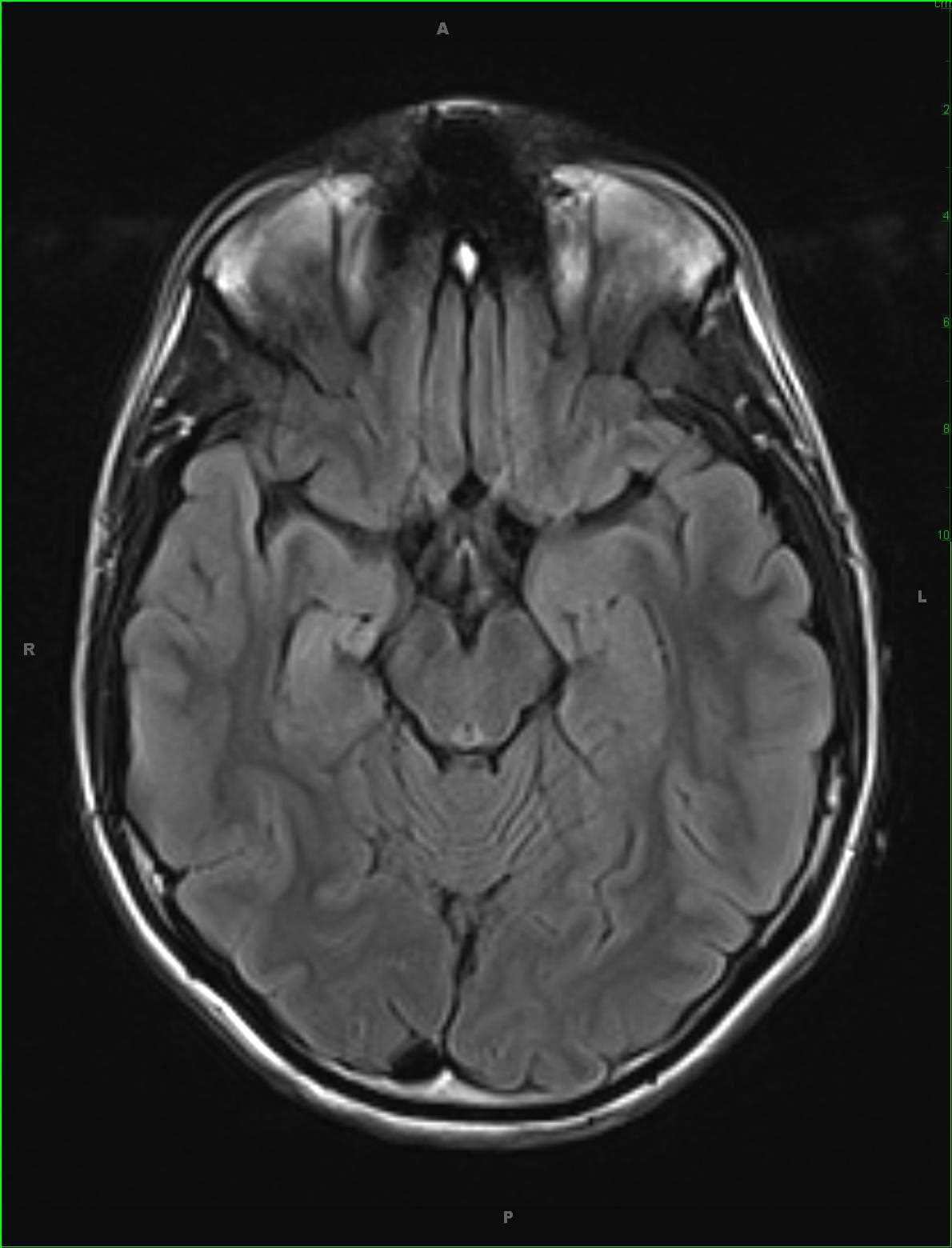
Mesial Temporal Sclerosis, Right
Note
28-year-old male with a long-standing history of seizures localizing to the inferomedial right temporal lobe on EEG. The hippocampus is small on the right with increased T2FLAIR signal intensity and a somewhat disorganized anatomic configuration of the cortical gray matter and subcortical white matter. In addition, the fornix is small on the right as compared to the left, and the right mamillary body is hypoplastic as compared to the left. There is slight dilatation of the right temporal horn as compared to the left. These imaging findings are compatible with mesial temporal sclerosis on the right. Mesial temporal sclerosis is the most common association with intractable temporal lobe epilepsy. Most patients present with complex partial temporal lobe seizures. On histologic sectioning, there is neuronal cell loss, gliosis, and sclerosis. Typical MR imaging findings include atrophy of the ipsilateral fornix and mammillary body, increased signal intensity and atrophy of the anterior thalamic nucleus, atrophy of the cingulate gyrus, reduction in volume of the amygdala, and reduction in volume of the subiculum among many other imaging features. Treatment is primarily with antiepileptic agents, but in those who are refractory to medical management, temporal lobectomy or selective amygdalohippocampectomy may be performed.
Related videos to the case