- 2
- ,
- 2
- 4
- 6
To Quiz Yourself: Select OFF by clicking the button to hide the diagnosis & additional resources under the case.
Quick Browser: Select ON by clicking the button to hide the additional resources for faster case review.
CASE NUMBER
277


Diagnosis
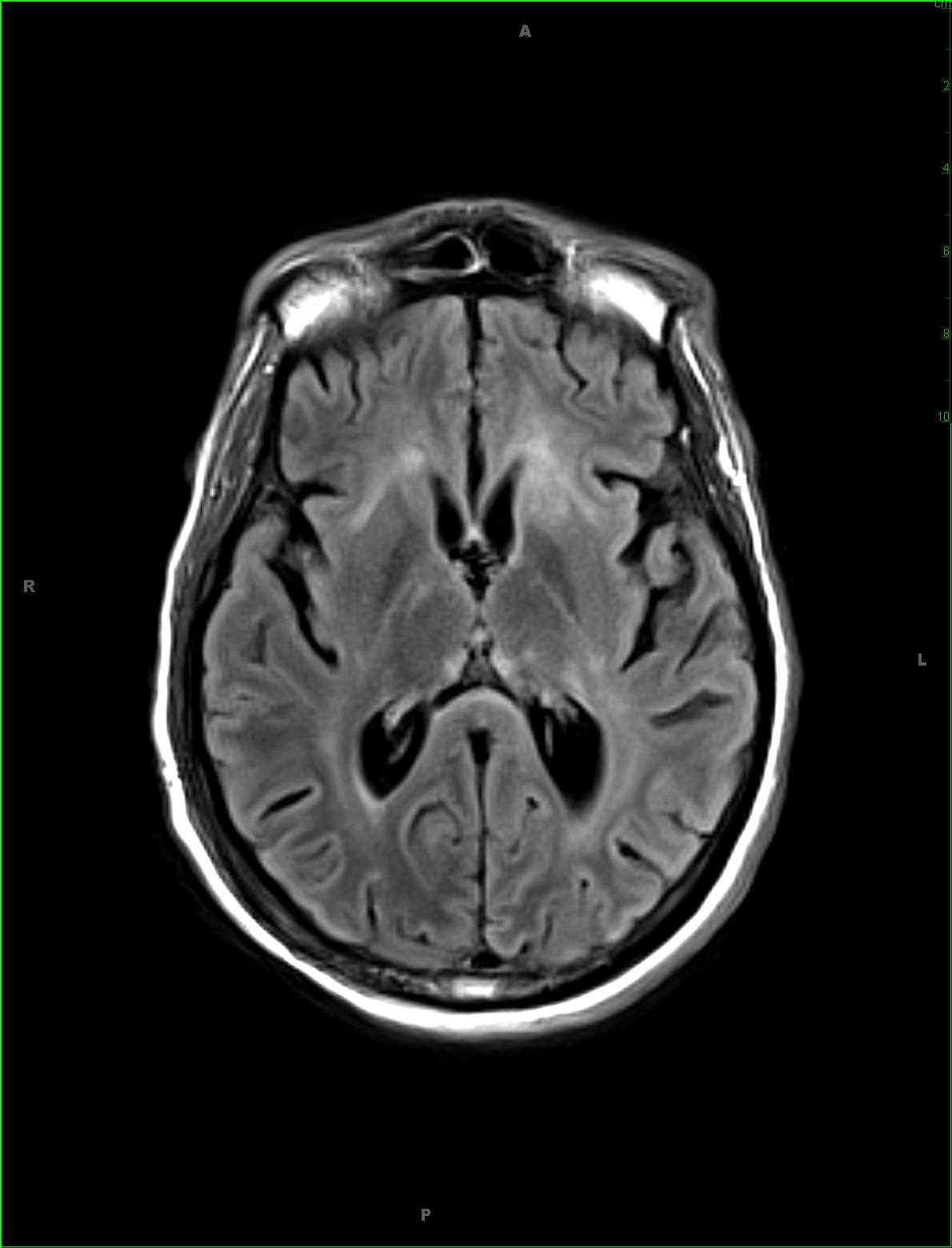
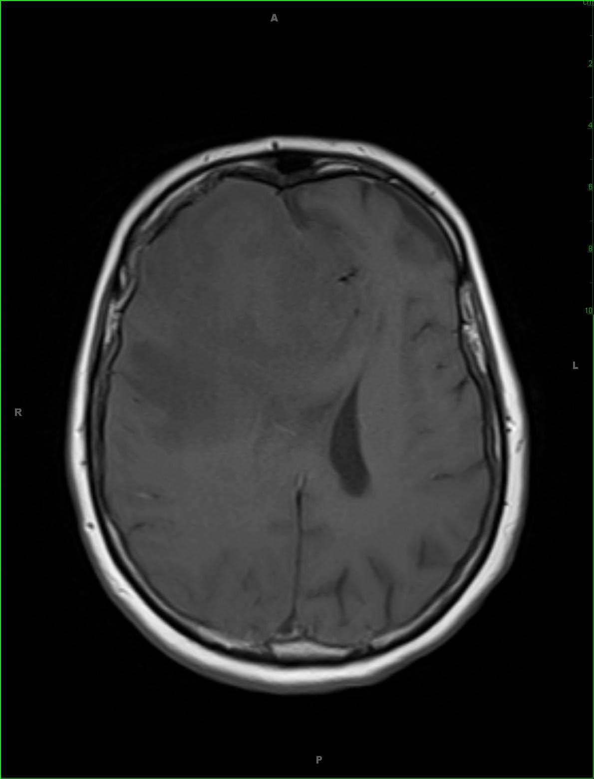
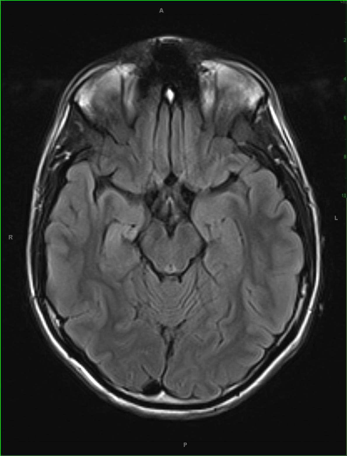
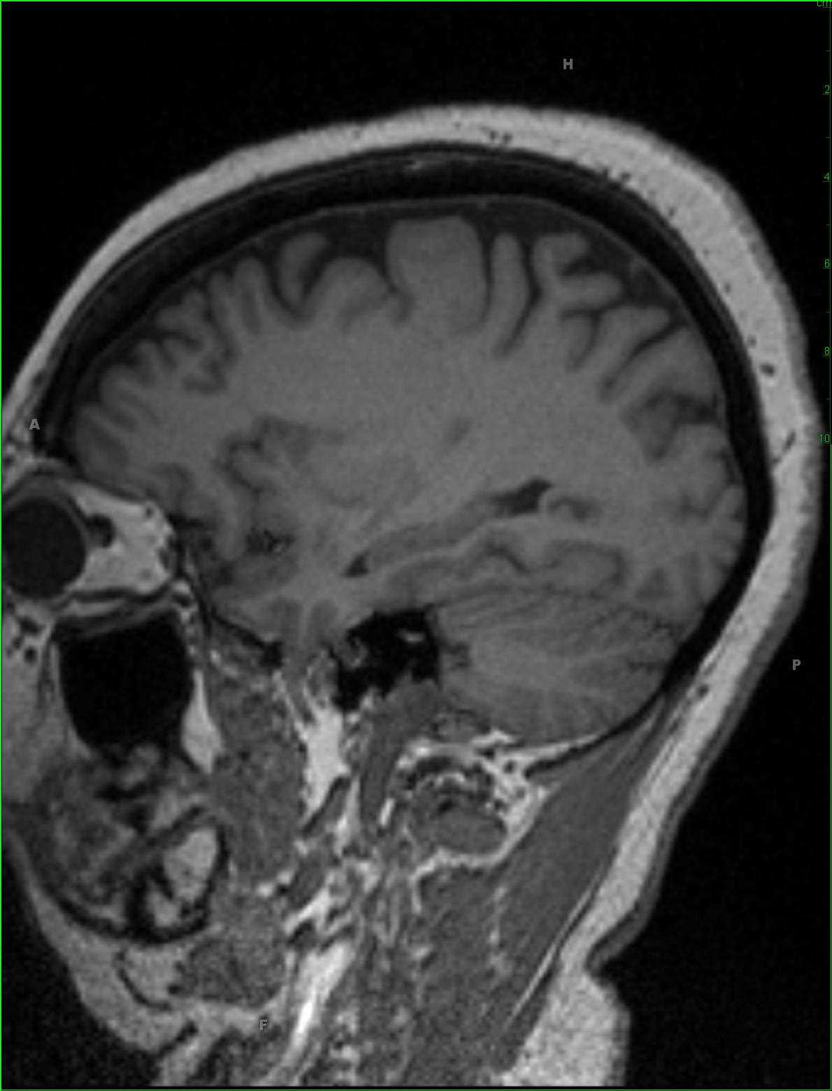
Inferior Right Temporal Encephalocele
Note
32-year-old male with a history of long-standing seizures localizing to the inferior right temporal region on EEG. Images demonstrate herniation of inferior right temporal lobe tissue with a somewhat tethered and distorted appearance into the skull base on the right at the level of the foramen ovale. These images depict an inferior right temporal encephalocele. Encephalocele should be distinguished from meningoceles which the lesion contains only meninges herniating through a dural and skull defect. Clinical presentation may range from an apparent subcutaneous mass at birth while skull base encephaloceles may present as a lump or bump in the oropharynx or nasopharynx. The lesions are thought to be secondary to failure of closure of the rostral end of the neural tube in early development. The tissue extending through the osseous defect is typically dysplastic and may be a source of recurrent seizures. Treatment is with resection of the dysplastic tissue and duraplasty at the osseous and dural defect.
Related videos to the case