- 2
- ,
- 2
- 4
- 6
To Quiz Yourself: Select OFF by clicking the button to hide the diagnosis & additional resources under the case.
Quick Browser: Select ON by clicking the button to hide the additional resources for faster case review.
CASE NUMBER
280


Diagnosis
Meningioma with Myxoid Features
Note
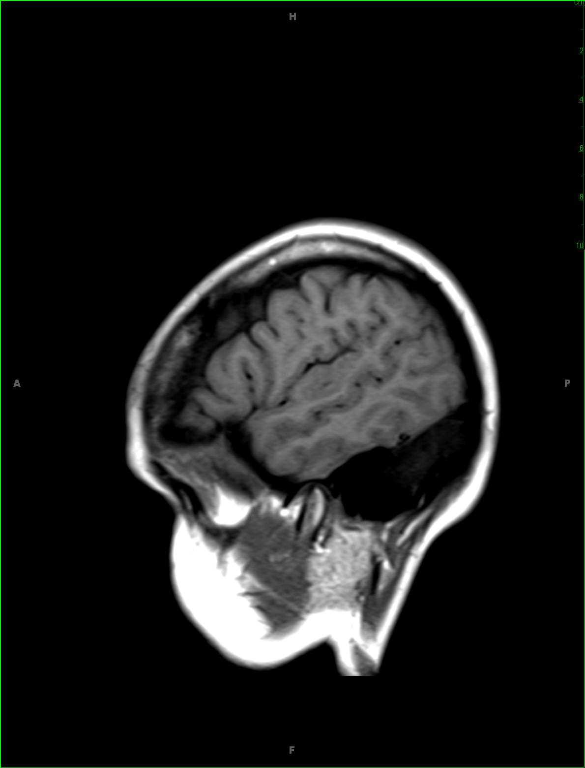
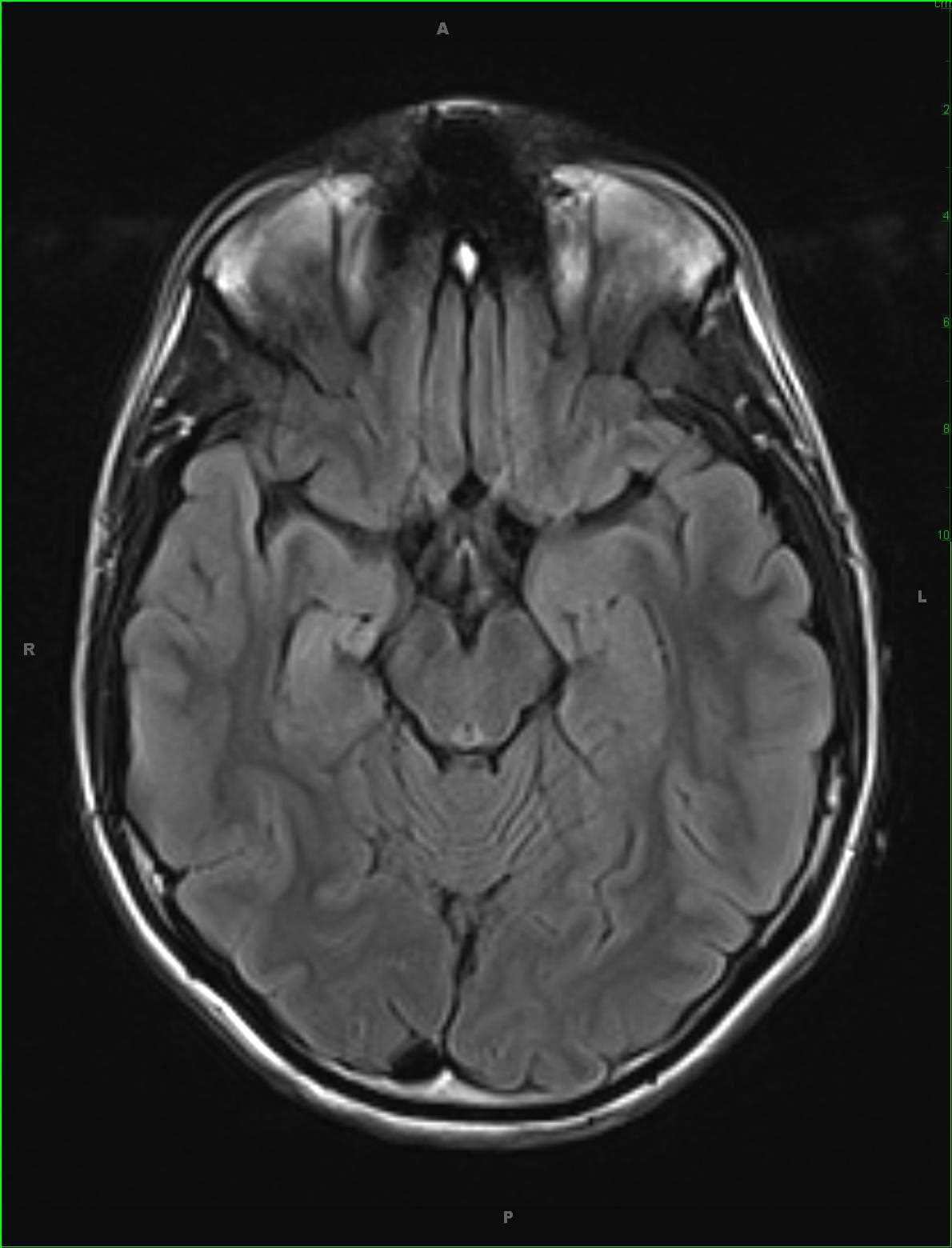
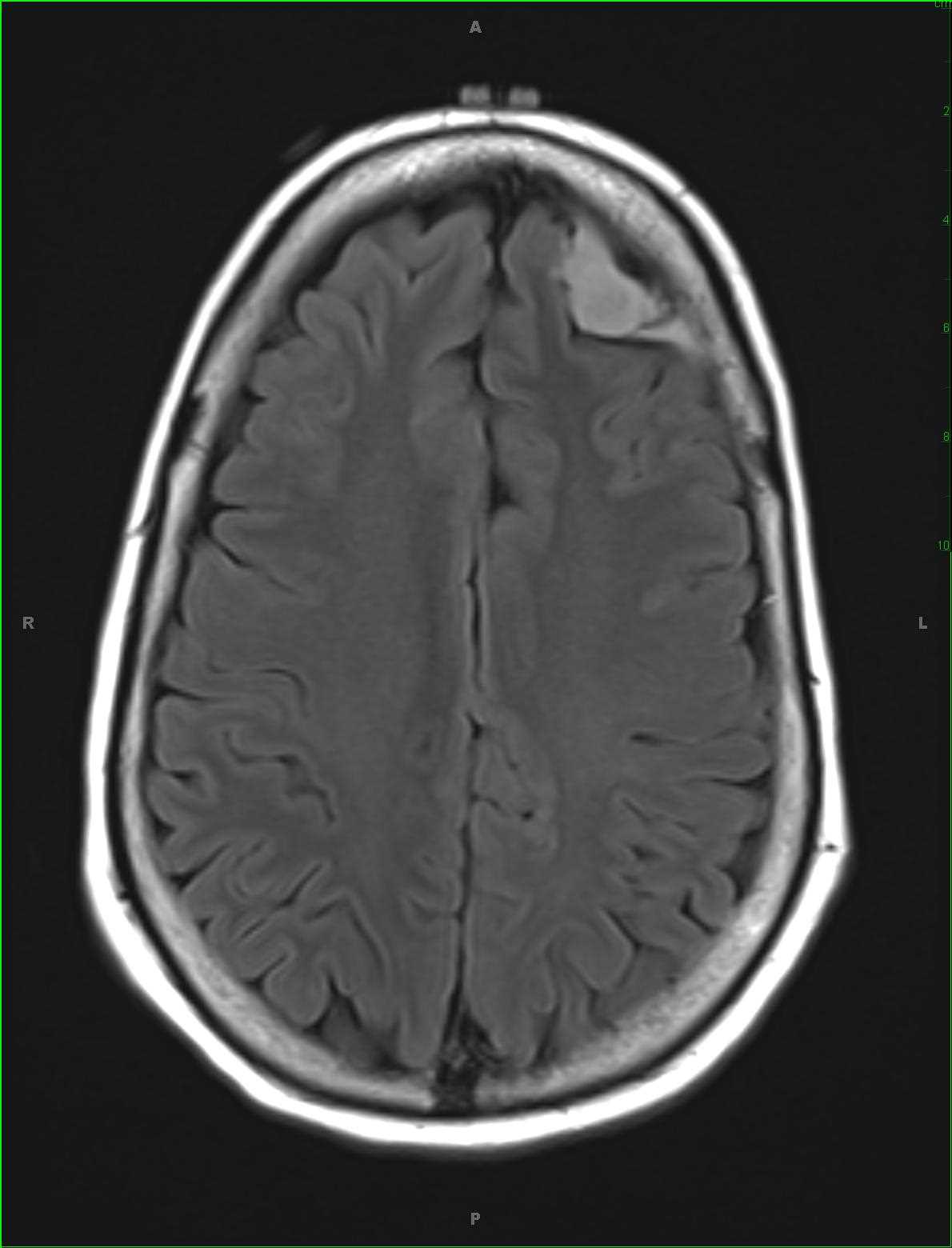
56-year-old female who underwent brain MRI examination for chronic headaches following an automobile accident in which an incidental extra-axial mass was identified over the left frontal region for which a preoperative MRI examination was ordered. The images demonstrate an extra-axial dural based T2/FLAIR hyperintense predominately solid lesion with a small hemorrhagic component along its posterolateral margin with more peripheral hemosiderin deposition and lobular T2/FLAIR hyperintense regions which restrict diffusion. There is prominent signal loss on the fat saturated T2 weighted images at that site. A large dural tail is identified on the postcontrast images. The imaging findings are most consistent with a meningioma. Following resection, a myxoid meningioma histopathologic diagnosis was given. Myxoid meningiomas do not display the usual histologic features of more typical meningiomas, and contain abundant mucoid stroma similar to other myxoid tumors. The differential includes other lesions such as schwannomas, fibromyxoma, or fibroxanthoma. Treatment is with primary surgical resection with recurrence rates similar to non-myxoid meningioma subtypes.
Related videos to the case


THIS IS CASE
280
OF
373