- 2
- ,
- 2
- 4
- 6
To Quiz Yourself: Select OFF by clicking the button to hide the diagnosis & additional resources under the case.
Quick Browser: Select ON by clicking the button to hide the additional resources for faster case review.
CASE NUMBER
259


Diagnosis
Metastatic Melanoma, with ciliary body (globe) involvement.
Note
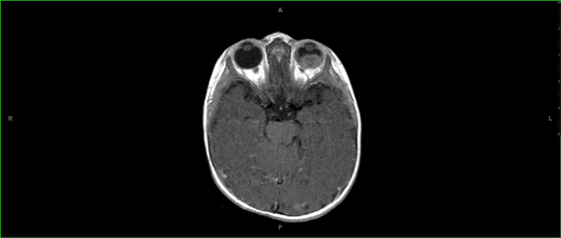
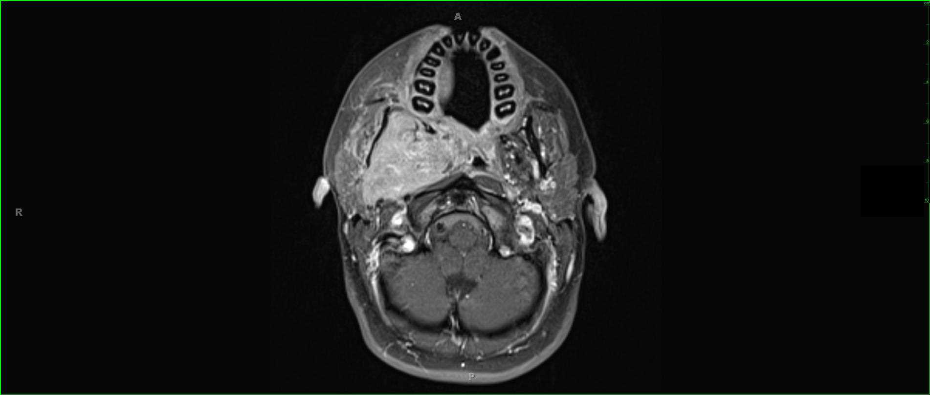
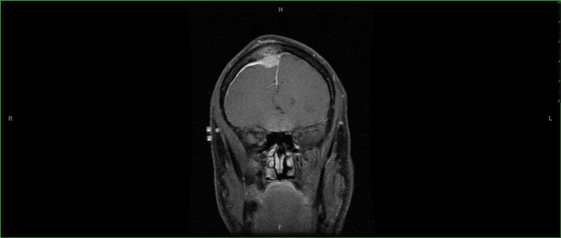
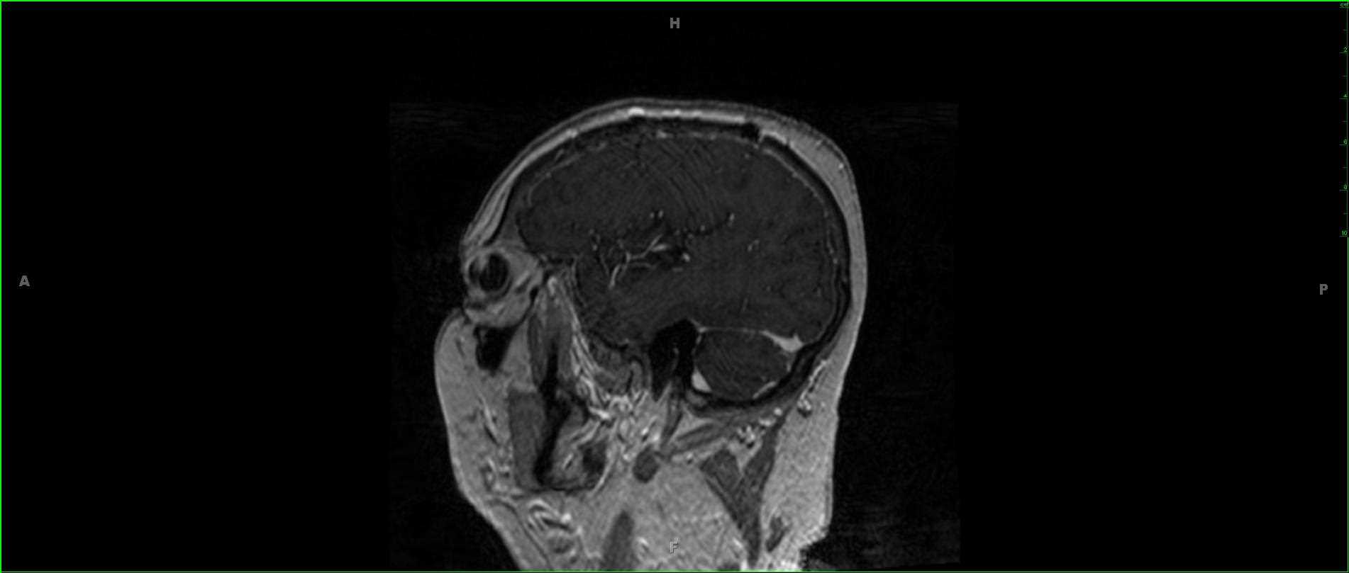
These images demonstrate 2 partially solid and partially cystic T1 hyperintense, enhancing, hemorrhagic lesions involving the left cerebral hemisphere in this 68-year-old female. A convex margin is identified along the superolateral aspect of the right vitreous. There is prominent T2 hypointense signal with enhancement. The patient had a primary diagnosis of metastatic melanoma including 2 metastatic lesions within the left cerebral hemisphere. A third lesion is identified centered in the ciliary body of the right orbit. Metastatic ocular disease to the globe is the second most common intraocular mass lesion in the adult population. The most common primary intraocular lesion in the adult population is primary uveal melanoma. Other causes include hemangioma, leiomyoma, and osteoma. The most common primary sites of metastaic disease to the globe include lung and breast cancer, though melanoma and renal cell carcinoma may also be seen. Retinal detachment may or may not be present.
Related videos to the case