- 2
- ,
- 2
- 4
- 6
To Quiz Yourself: Select OFF by clicking the button to hide the diagnosis & additional resources under the case.
Quick Browser: Select ON by clicking the button to hide the additional resources for faster case review.
CASE NUMBER
260


Diagnosis
Intra-osseous Meningioma and Subgaleal Invasion
Note
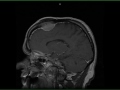
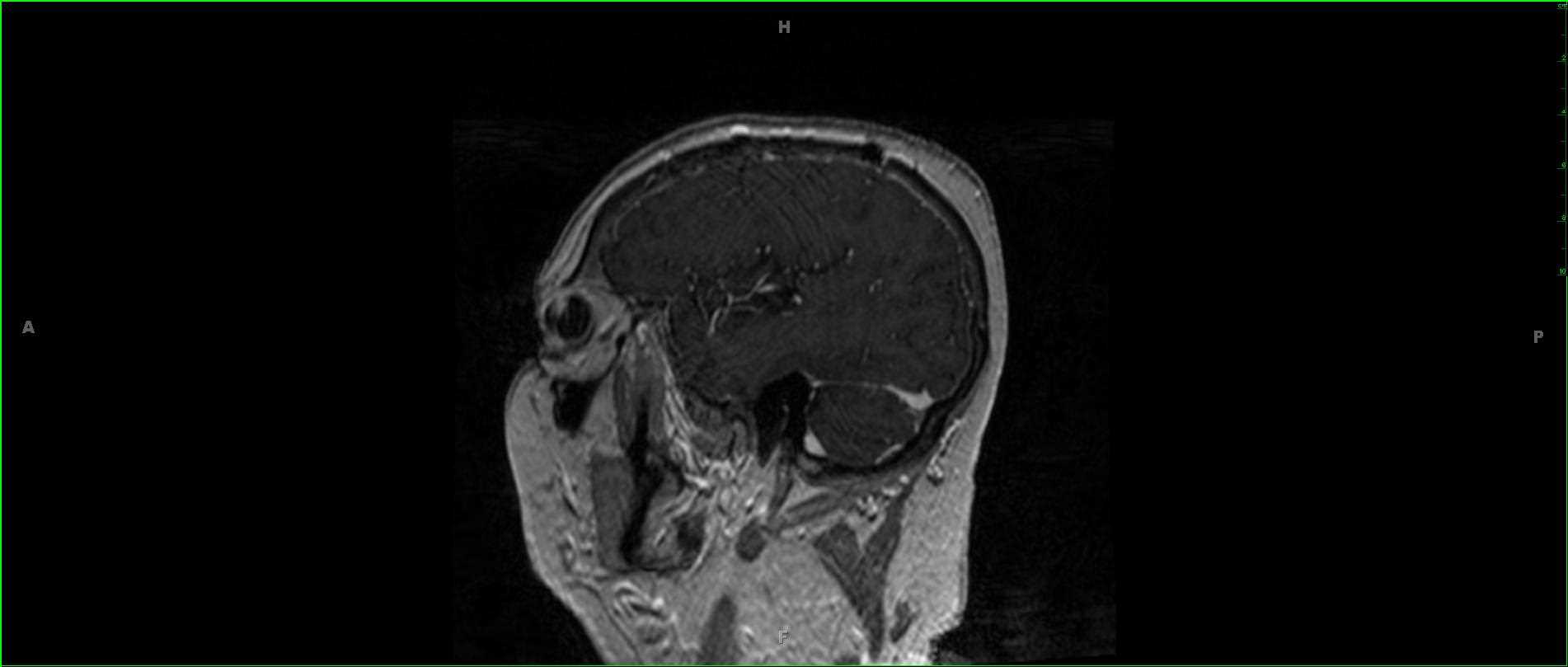
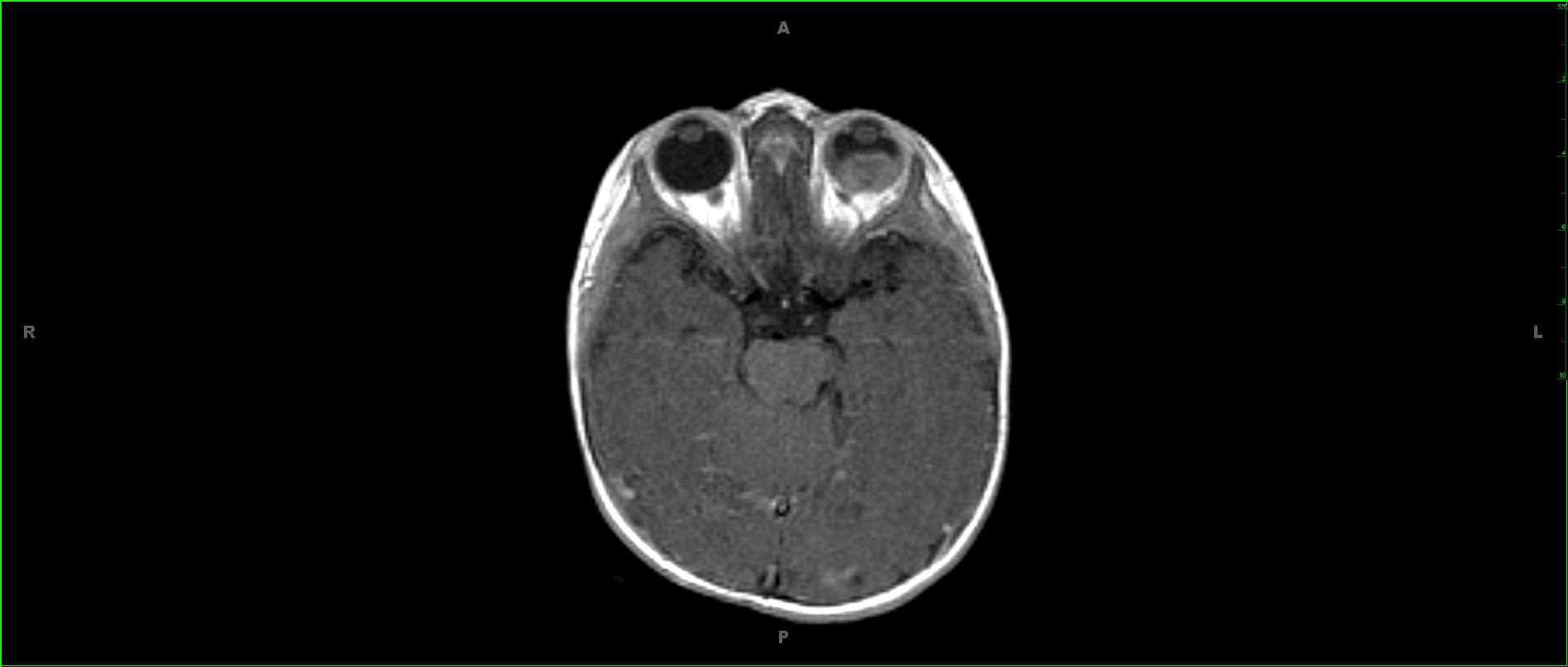
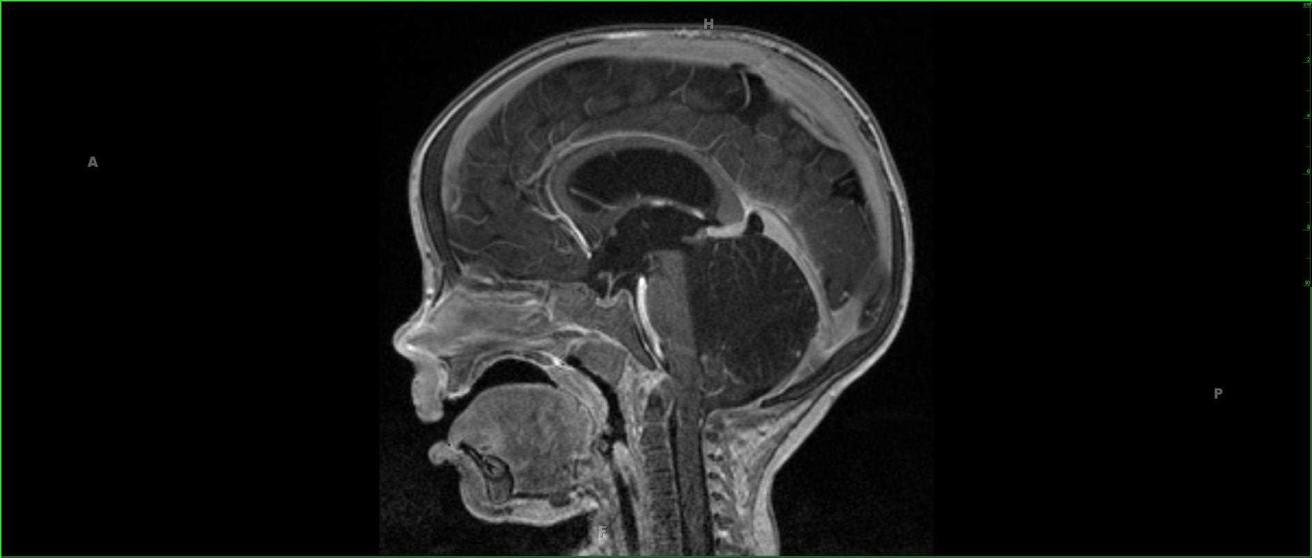
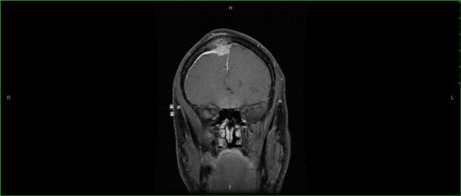
72-year-old male who presented with chronic headaches and a small lump in the right frontal scalp region near the midline. There is a partially circumscribed infiltrative, T1 hypointense, FLAIR minimally hyperintense mass centered in the right frontal region. The lesion demonstrates moderate diffusion restriction, and there is enhancing tissue extending into and through the right frontal bone to the subgaleal space. There is associated linear dural thickening. The patient had a history of prostate adenocarcinoma, and a differential was given which includes metastatic disease, hemangiopericytoma, or less likely meningioma. On resection, this was found to be a meningioma with intraosseous and subgaleal extension. Intra-osseous meningiomas are a rare subtype of meningioma accounting for less than 1% of osseous tumors. Clinical presentation is usually due to mass effect on surrounding structures. Lesions are thought to arise from trapped arachnoid meningeal cap cells within the cranial sutures during development. There is a slightly higher percentage of malignant change when compared to standard meningiomas with surgical resection and bone grafting typically performed as treatment.
Related videos to the case