- 2
- ,
- 2
- 4
- 6
To Quiz Yourself: Select OFF by clicking the button to hide the diagnosis & additional resources under the case.
Quick Browser: Select ON by clicking the button to hide the additional resources for faster case review.
CASE NUMBER
258


Diagnosis
Retinoblastoma
Note
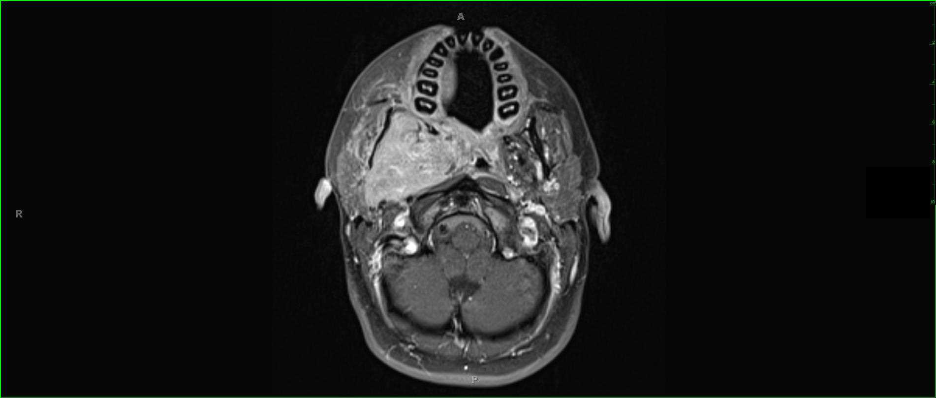
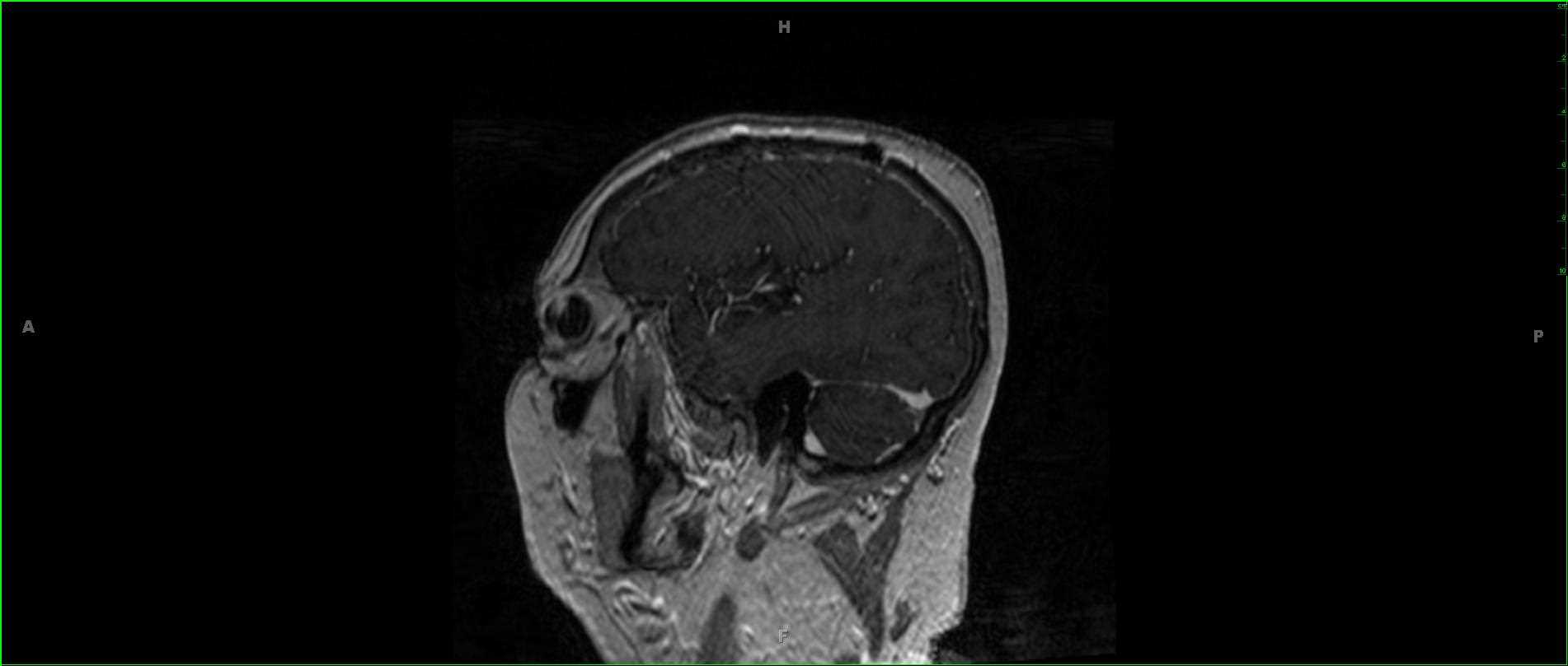
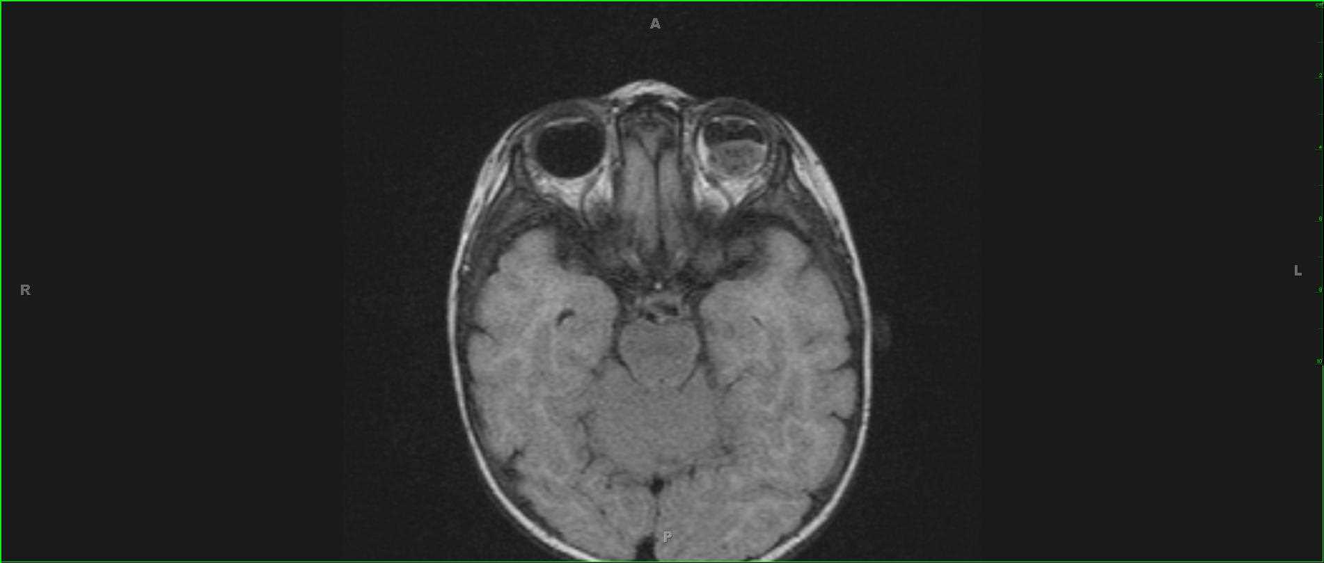
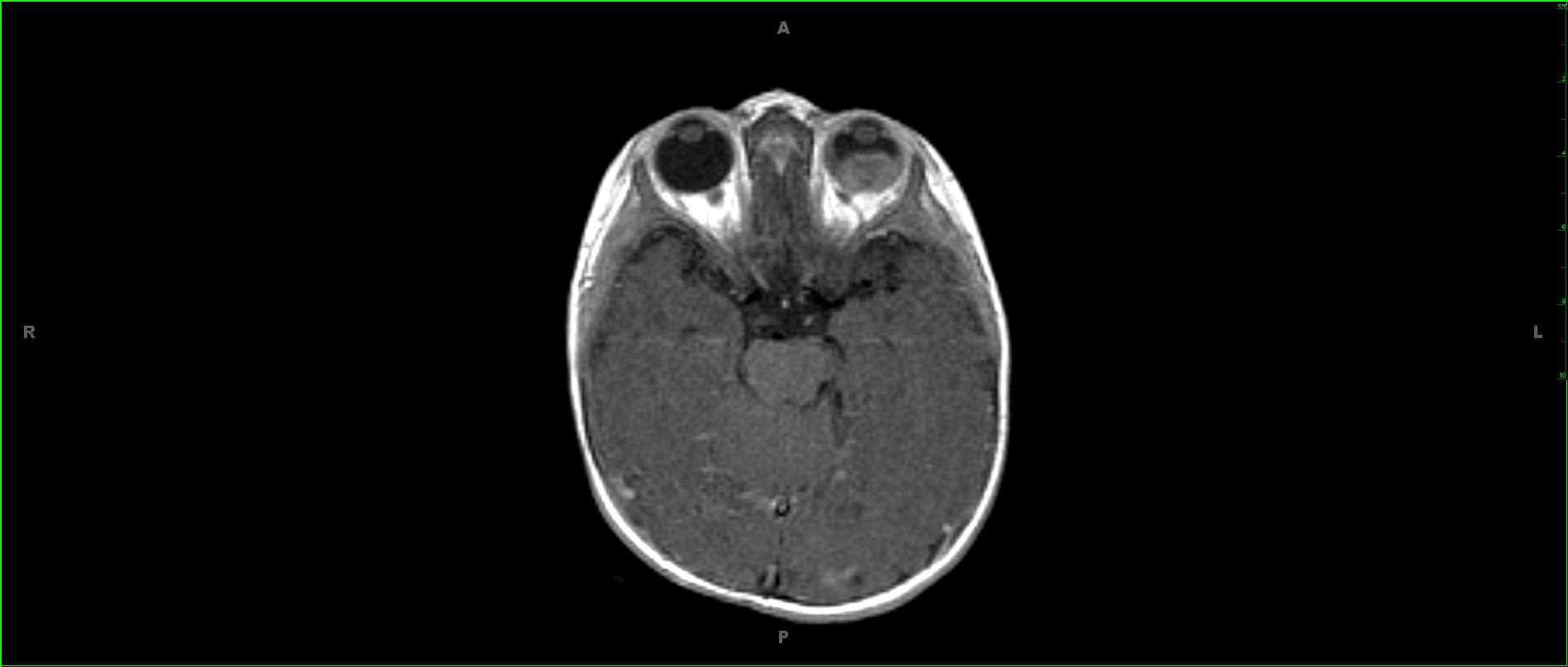
8-year-old male with leukocoria on the left. These images demonstrate a globular, T1 slightly hypointense, T2 heterogeneously slightly hypointense mass centered within the globe posteriorly on the left. The lesion is somewhat speckled in appearance with foci of signal loss on both the T1 and fluid sensitive images representing focal mineralization/calcifications. The diffusion weighted images demonstrate prominent restriction. There is secondary retinal detachment with subretinal hemorrhage. The lesion demonstrates mildly heterogeneous enhancement. This is a case of retinoblastoma on the left. Retinoblastoma is the most common intraocular tumor in childhood. 90% of cases are sporadic. Retinoblastomas are tumors of neuroectodermal cells which become retinal photoreceptors under normal conditions. The most common clinical presentation is leukocoria followed by strabismus. Bilaterality occurs in approximately 30% of patients affected by the hereditary form of disease.