- 2
- ,
- 2
- 4
- 6
To Quiz Yourself: Select OFF by clicking the button to hide the diagnosis & additional resources under the case.
Quick Browser: Select ON by clicking the button to hide the additional resources for faster case review.
CASE NUMBER
206


Diagnosis
Neurofibromatosis Type 1
Note
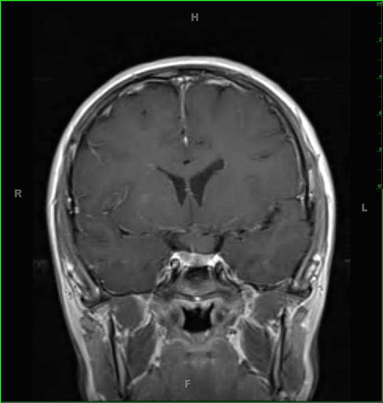
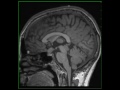
25-year-old female with a history of neurofibromatosis type 1 (NF-1). There are numerous poorly circumscribed FLAIR-hyperintense lesion in the basal ganglia as well as the dentate nuclei region bilaterally. There is nodular enlargement of the optic chiasm and prechiasmatic segment of the optic nerve on the left. The enlarged optic nerve contacts but remains distinct from the infindibulum of the pituitary gland, as is demonstrated on the postcontrast images. The findings are compatible with an optic pathway glioma in the setting of NF-1. The patient also had numerous subcutaneous neurofibromas in the suboccipital region (not demonstrated on these images). NF-1 is also known as von Recklinghausen disease, which is the most common phakomatosis. 50% of cases are due to new mutations while the other half are due to inherited autosomal dominant mutations. To make the clinical diagnosis, two or more of the following are required: > 6 cafe au lait spots, 2+ neurofibromas or one plexiform neurofibroma, optic nerve glioma, osseous lesions (ribbon ribs, gracile bones, multiple non-ossifying fibromas, etc), sphenoid wing dysplasia, 2+ iris hamartomas (Lisch nodules), axillary or inguinal freckling and primary relative with NF-1.
Related videos to the case

THIS IS CASE
206
OF
373