- 2
- ,
- 2
- 4
- 6
To Quiz Yourself: Select OFF by clicking the button to hide the diagnosis & additional resources under the case.
Quick Browser: Select ON by clicking the button to hide the additional resources for faster case review.
CASE NUMBER
139


Diagnosis
Cavernoma
Note
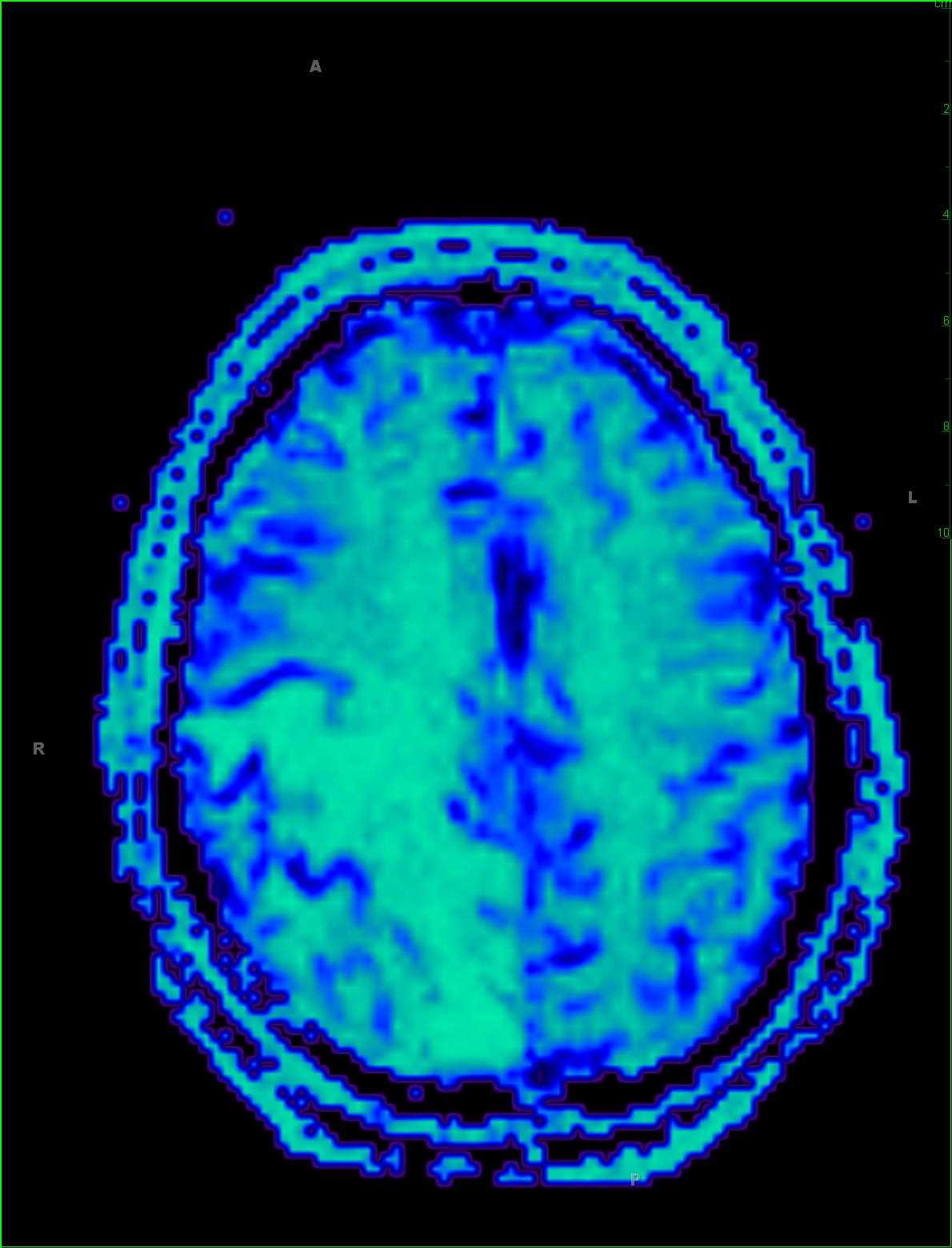
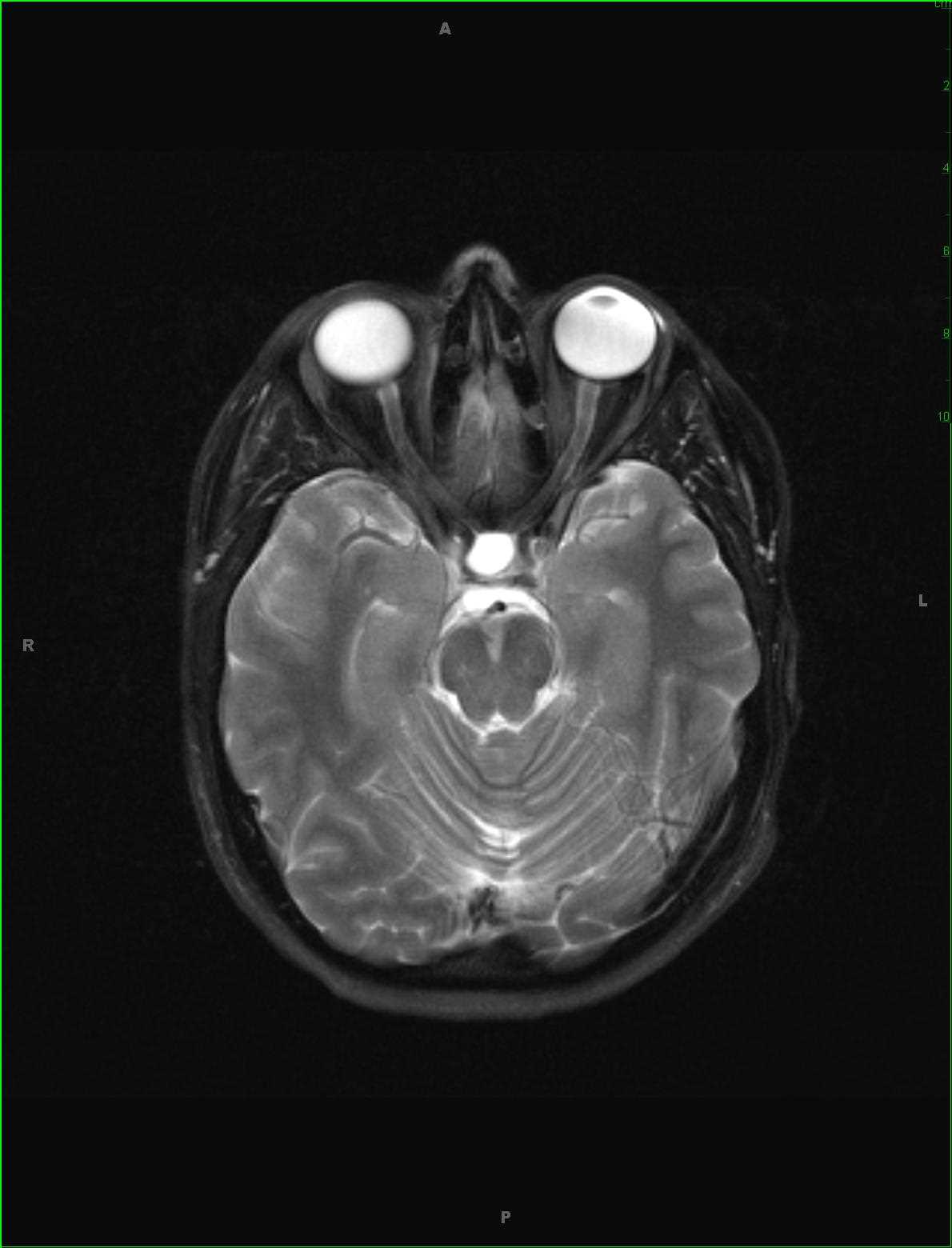
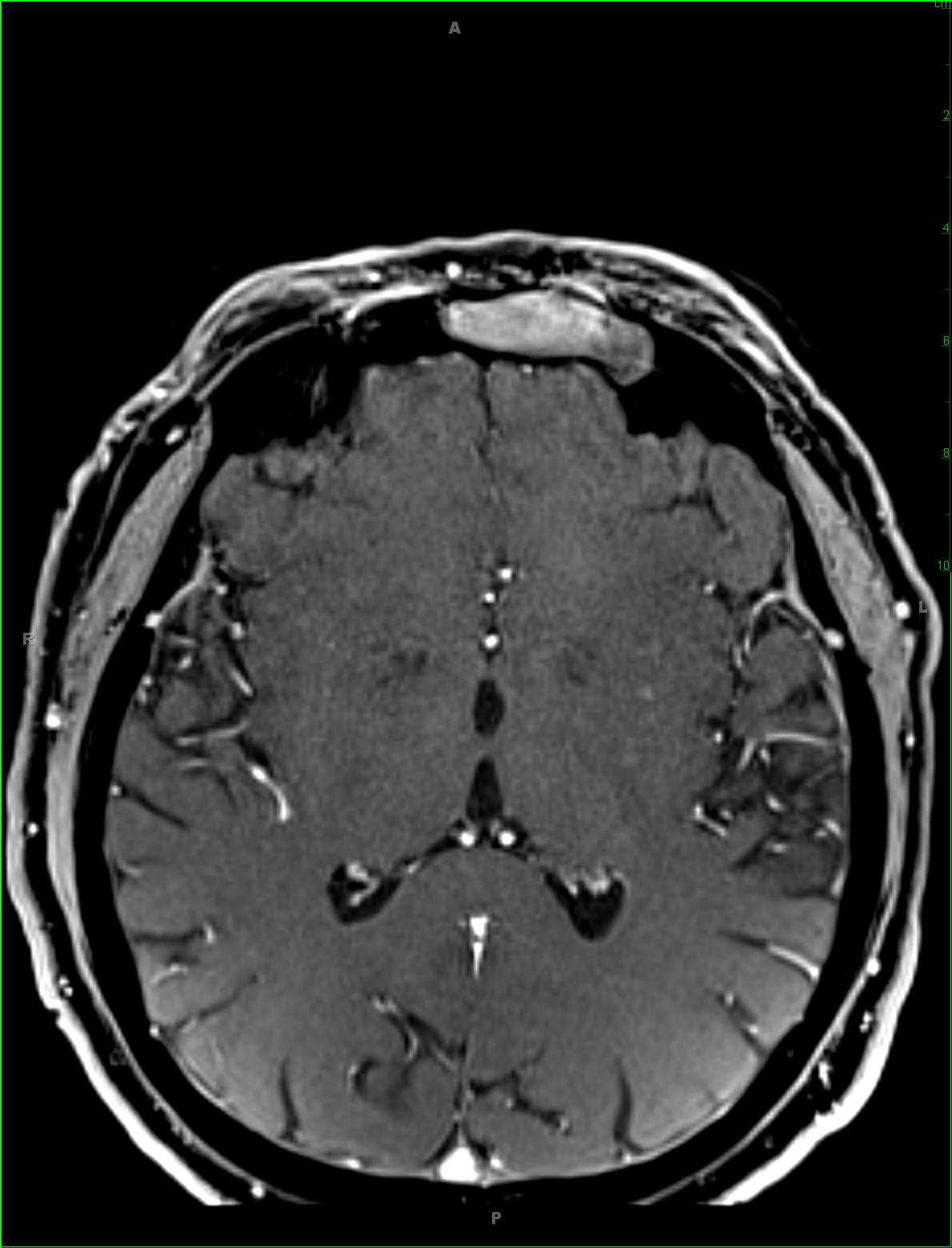
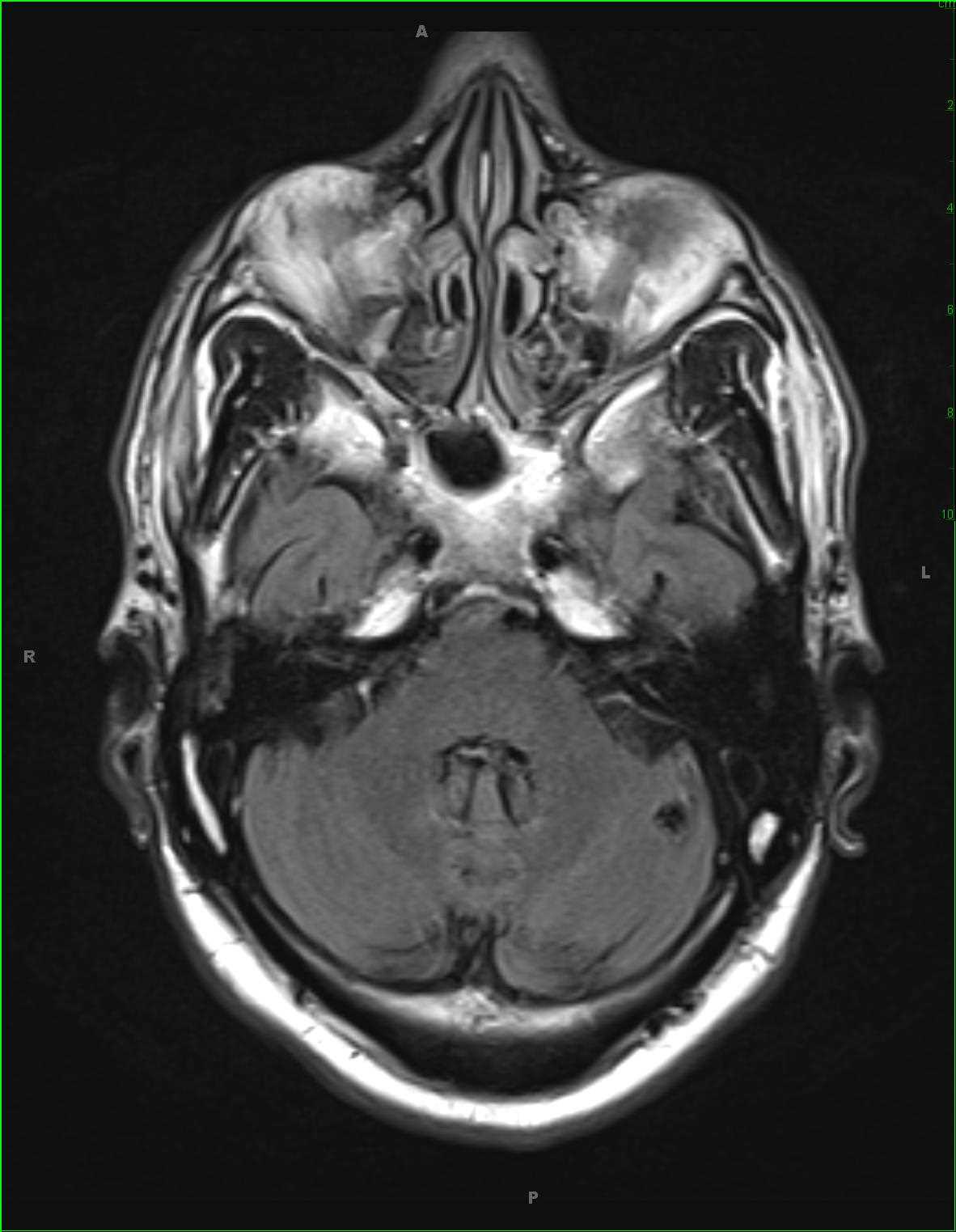
This is a 26-year-old female wo presented for new seizure-like activity. There is a circumscribed subcentimeter hypointense lesion demonstrated on images 1-5, the T2, DWI, ADC map, SWI, and FLAIR-weighted sequences, respectively. The final image, a noncontrast 3D-axial T1-weighted sequence, demonstrates a tiny central focus of T1-isointense signal. The patient also demonstrated several additional hypointense lesions throughout the supratentorial compartment. These imaging findings are classic for a small cavernoma, without recent hemorrhage. MR imaging features include a lesion with a hypointense rim on all sequences, but best visualized on the susceptibility weighted images. If hemorrhage is present, depending on the stage of acuity, the intrinsic T1- and T2-weighted signal is variable, resulting in the classic popcorn appearance. Cavernous malformations can be congenital or acquired. Multiple cavernous occur in about 20% of cases.
Related videos to the case


THIS IS CASE
139
OF
373