- 2
- ,
- 2
- 4
- 6
To Quiz Yourself: Select OFF by clicking the button to hide the diagnosis & additional resources under the case.
Quick Browser: Select ON by clicking the button to hide the additional resources for faster case review.
CASE NUMBER
138


Diagnosis
Hemorrhagic Infarct Secondary to Dural Sinus Thrombosis
Note
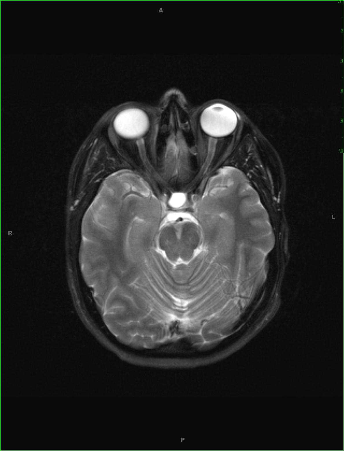
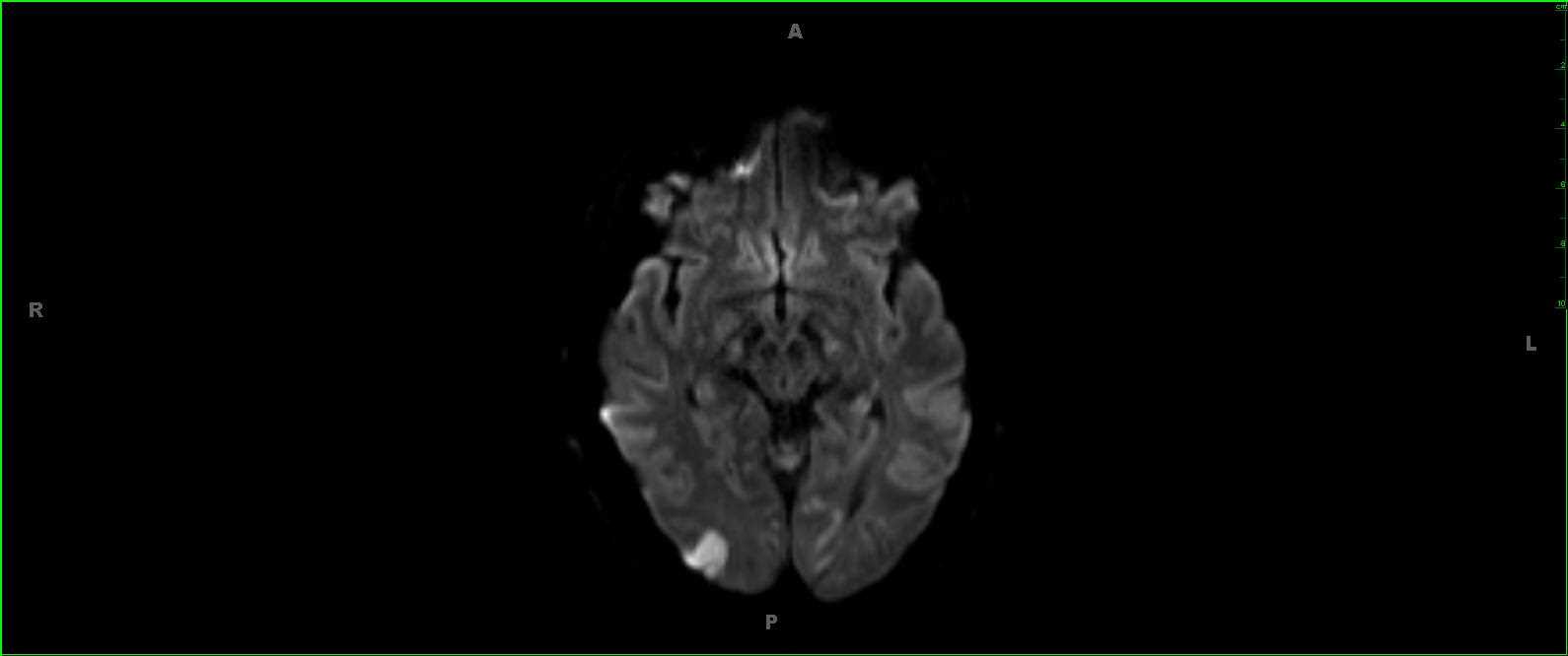
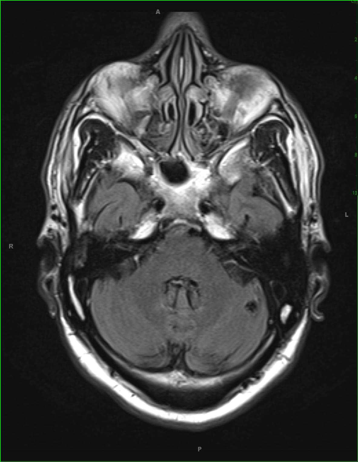
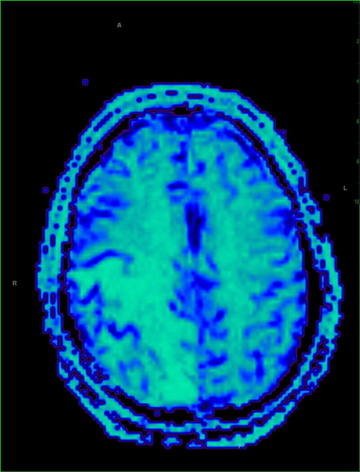
This is a case of venous hemorrhagic infarction secondary to dural venous sinus thrombosis in a 58-year-old female. A focal region of acute infarction with florid peripheral vasogenic edema is identified within the high right parietal lobe on the DWI and ADC maps, images 1 and 2. In addition, there are more focal regions of cytotoxic edema in the corona radiata bilaterally. Within a few of the sulci of the left parietal region, there is reduced diffusivity compatible with a small volume of subarachnoid hemorrhage at that site. Image 3, an axial T2/FLAIR-sequence, redemonstrates the florid edema involving the right > left parietal lobes with additional involvement of the posterior right frontal lobe. A few linear regions of signal loss are identified within the sulci of the biparietal area suspicious for subarachnoid hemorrhage at those sites. The presence and extent of the subarachnoid hemorrhage and hemorrhagic infarct is better assessed on susceptibility weighted image #4. The last two images demonstrate prolonged transit time in the right frontoparietal region due to venous congestion with a more focal region of reduced blood volume due to infarction. Patients with venous infarction demonstrate a variable clinical presentation dependent on cause, location, extent, rate of progression or regression, and collateral pathways to venous drainage.
Related videos to the case


THIS IS CASE
138
OF
373