- 2
- ,
- 2
- 4
- 6
To Quiz Yourself: Select OFF by clicking the button to hide the diagnosis & additional resources under the case.
Quick Browser: Select ON by clicking the button to hide the additional resources for faster case review.
CASE NUMBER
137


Diagnosis
Rathke's Cleft Cyst
Note
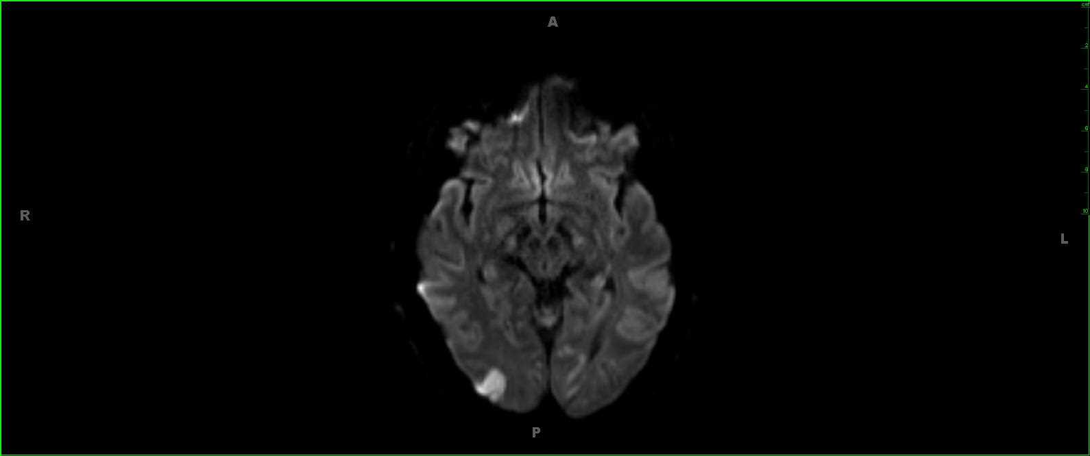
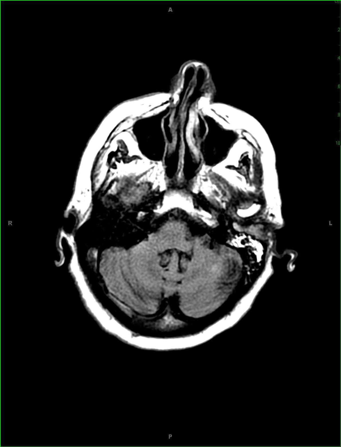
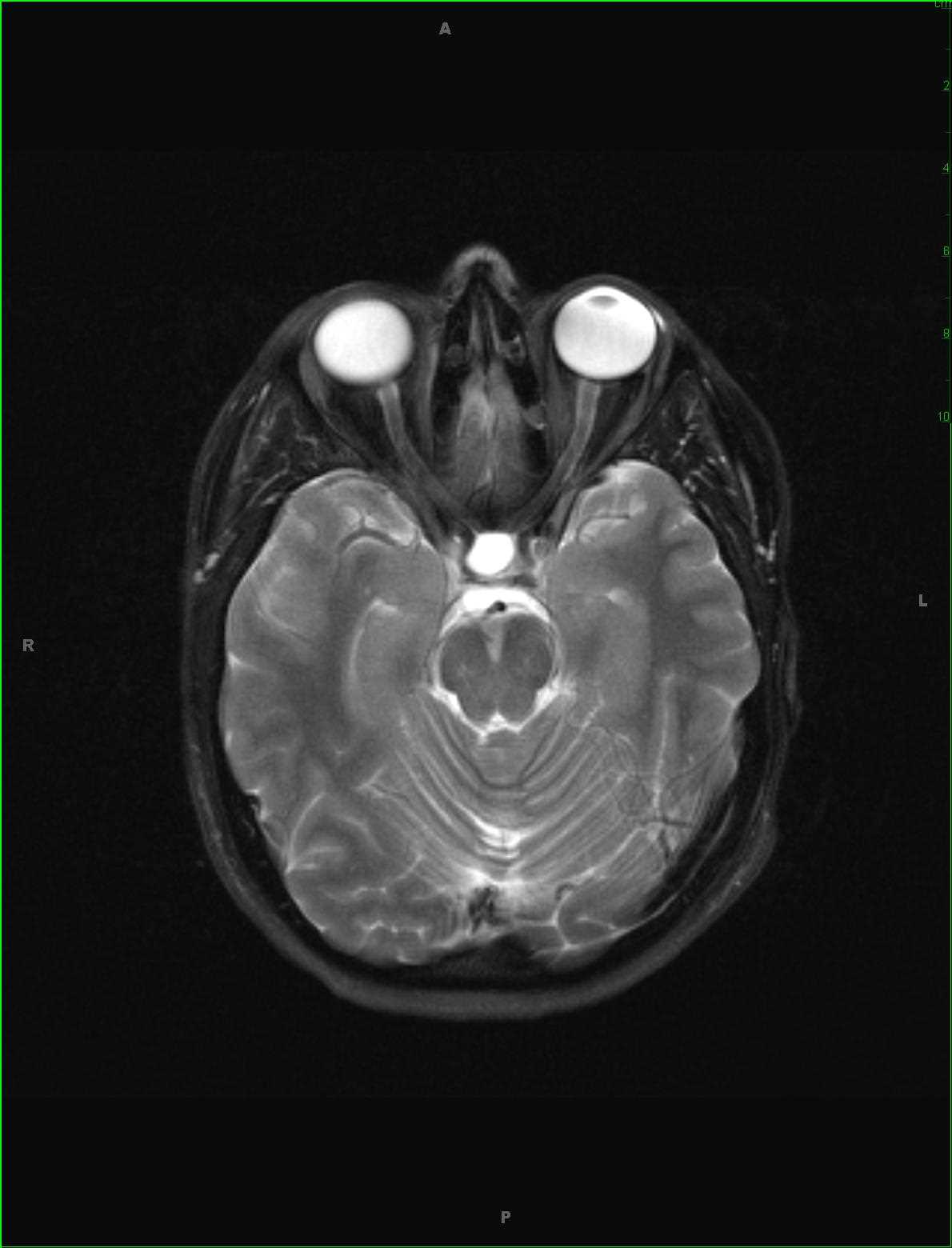
This is a case of a Rathkes cleft cyst in a 32-year-old female presenting with bitemporal hemianopsia. Images 1 and 2 demonstrate a well circumscribed, round, mildly T1-hypointense mass arising within and expanding the sella turcica. There is upward bowing of the diaphragma sella and contact with the inferior surface of the optic chiasm. The third image demonstrates a FLAIR hyperintense mass. The hyperintense signal and cystic nature of the lesion are redemonstrated on the axial T2-weighted image #4. Images 5 and 6 demonstrate no suspicious postcontrast enhancement. A Rathkes cleft cyst is a benign lesion originating as a remnant of Rathkes pouch. T1- and T2-weighted signal varies depending on the cyst contents (+/- mucin). The differential includes craniopharyngioma and pars intermedia cyst. There should be no septated or mural nodular enhancement in the setting of a Rathkes cleft cyst.
Related videos to the case