- 2
- ,
- 2
- 4
- 6
To Quiz Yourself: Select OFF by clicking the button to hide the diagnosis & additional resources under the case.
Quick Browser: Select ON by clicking the button to hide the additional resources for faster case review.
CASE NUMBER
140


Diagnosis
Mucocele
Note
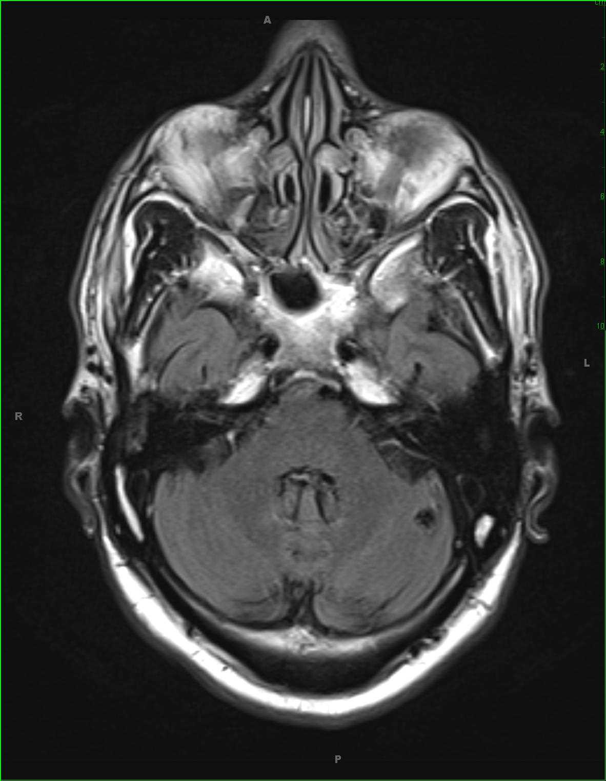
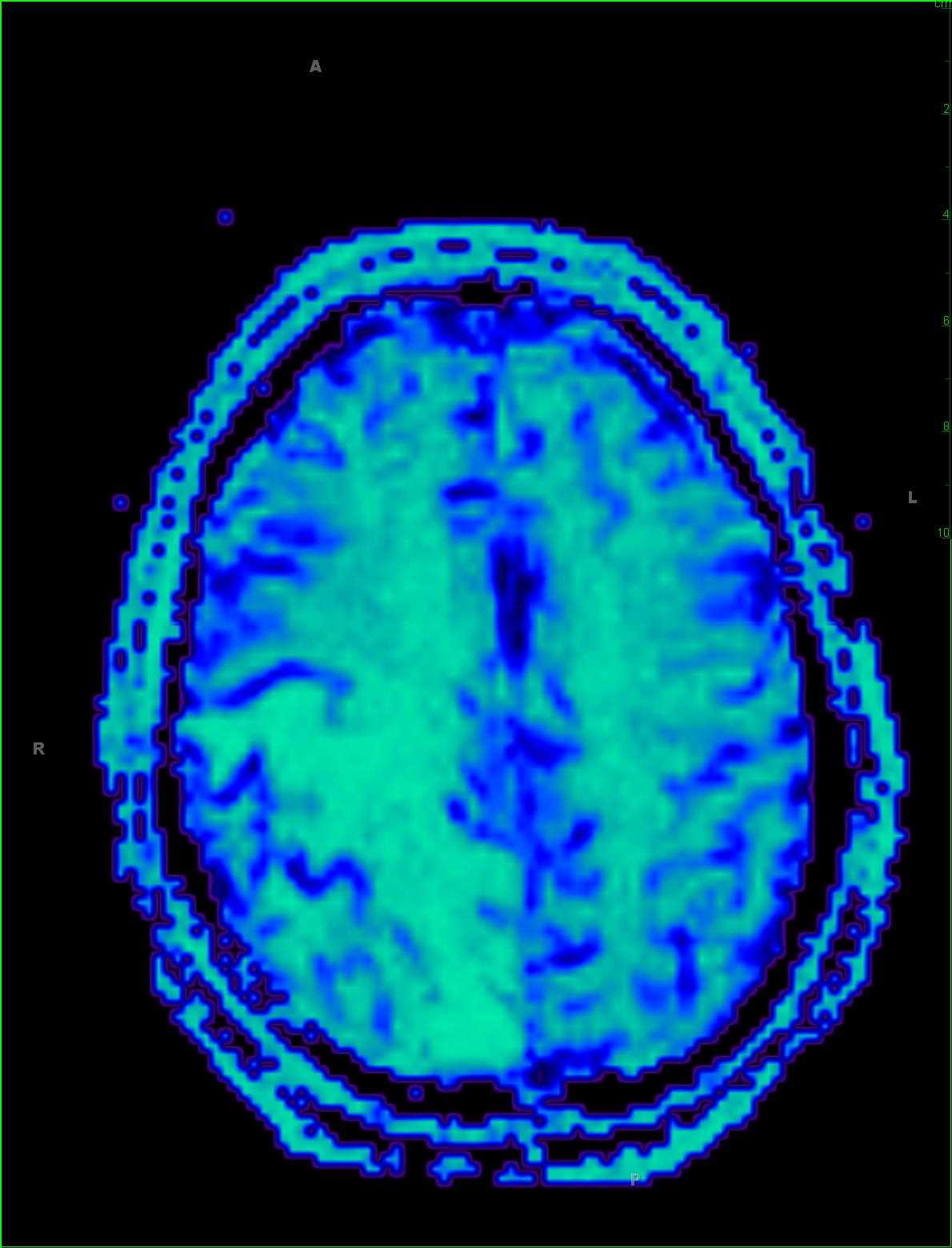
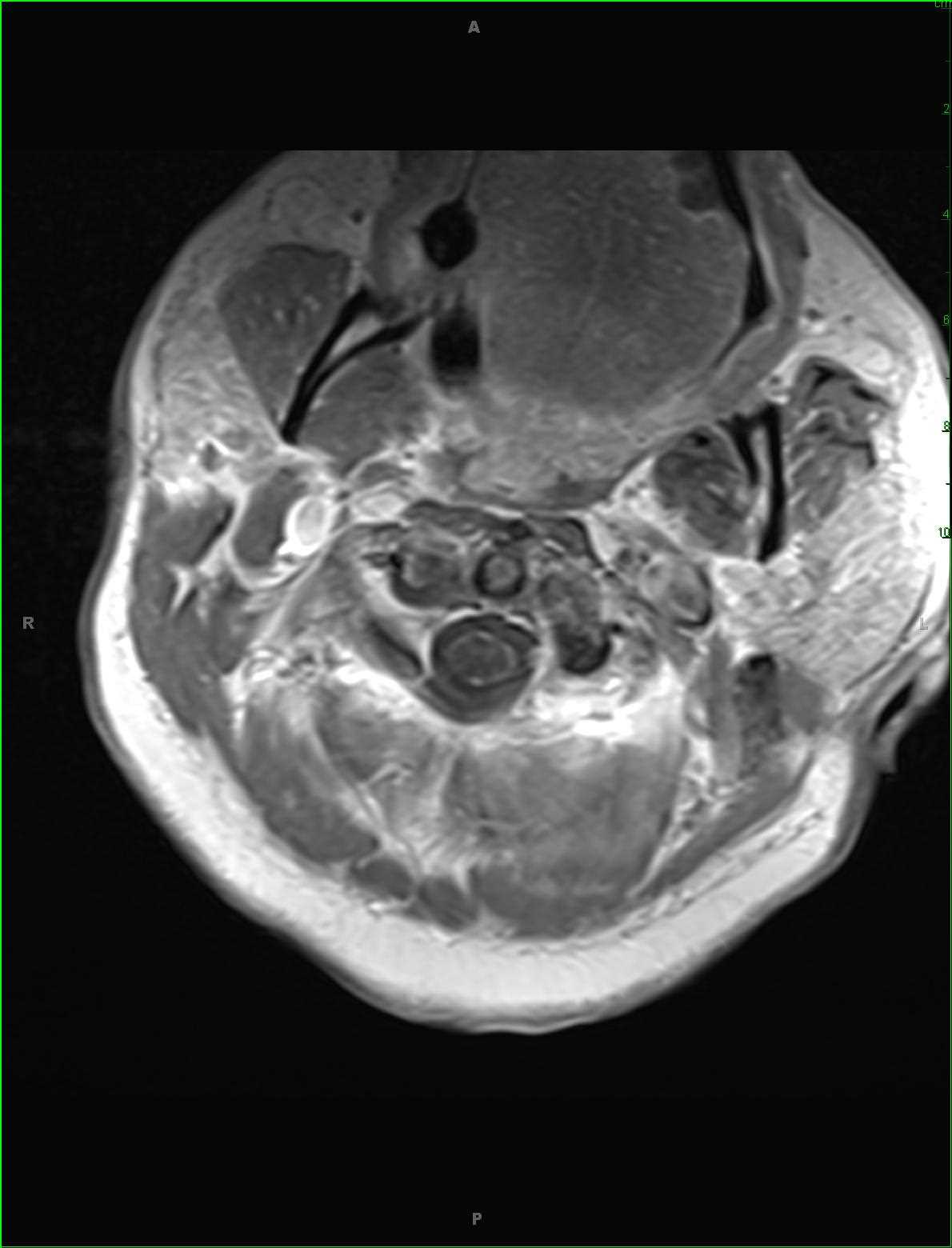
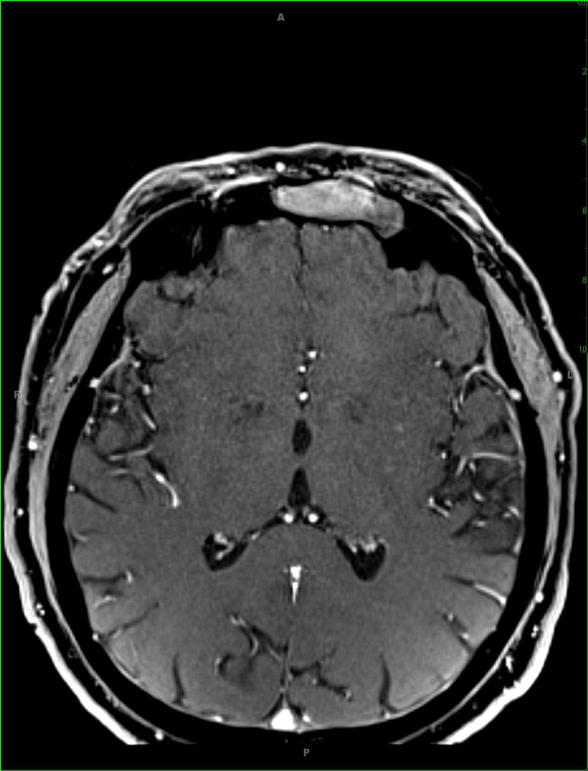
This is a case of a left frontal sinus mucocele in a 45-year-old male presenting with signs and symptoms of sinusitis for > 1 year. Image 1, a noncontrast axial T1-weighted sequence, demonstrates enlargement of the left frontal sinus with regions of intrinsic T1-hyper and hypointense signal. Image 2, an axial FLAIR sequence, demonstrates circumferential mucosal thickening, greatest laterally, with more FLAIR isointense signal within the central/medial left frontal sinus. The DWI and ADC maps, on images 3 and 4, demonstrate focal regions of reduced diffusivity within the medial aspect of the left frontal sinus. The fat-saturated axial T2 and post contrast fat-saturated axial T1-weighted sequences, images 5 and 6, redemonstrates the circumferential mucosal thickening and intrinsic T1-hyperintense material medially.These findings are classic for a left frontal sinus mucocele, which is the most common masslike complication of sinusitis. The variable protein content within the mucocele leads to variable signal intensity on the T1- and T2-weighted sequences. The most common sites of involvement are the frontal sinuses, ethmoid air cells, maxillary antra and sphenoid sinuses, in decreasing order of frequency.
Related videos to the case


THIS IS CASE
140
OF
373