- 2
- ,
- 2
- 4
- 6
To Quiz Yourself: Select OFF by clicking the button to hide the diagnosis & additional resources under the case.
Quick Browser: Select ON by clicking the button to hide the additional resources for faster case review.
CASE NUMBER
135


Diagnosis
External Auditory Canal Adenoid Cystic Carcinoma
Note
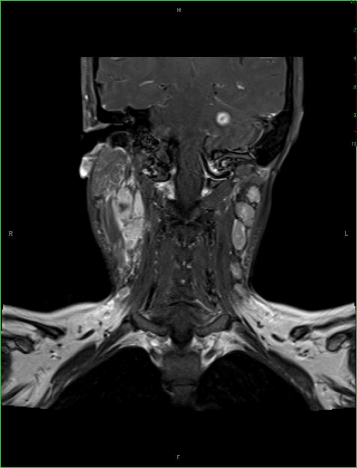
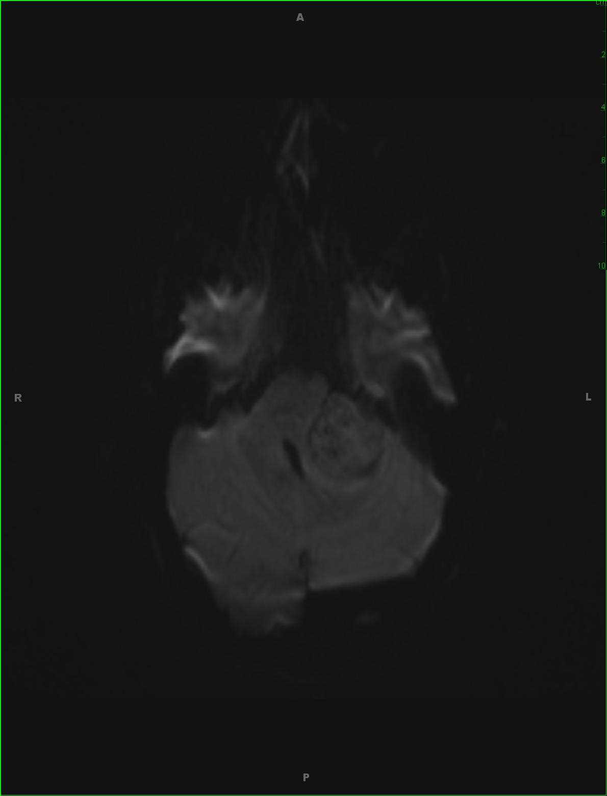
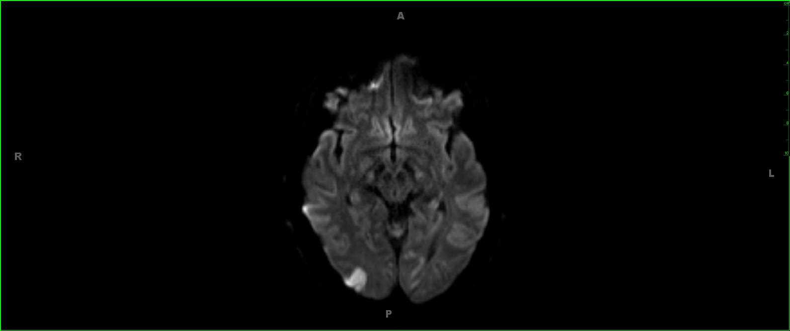
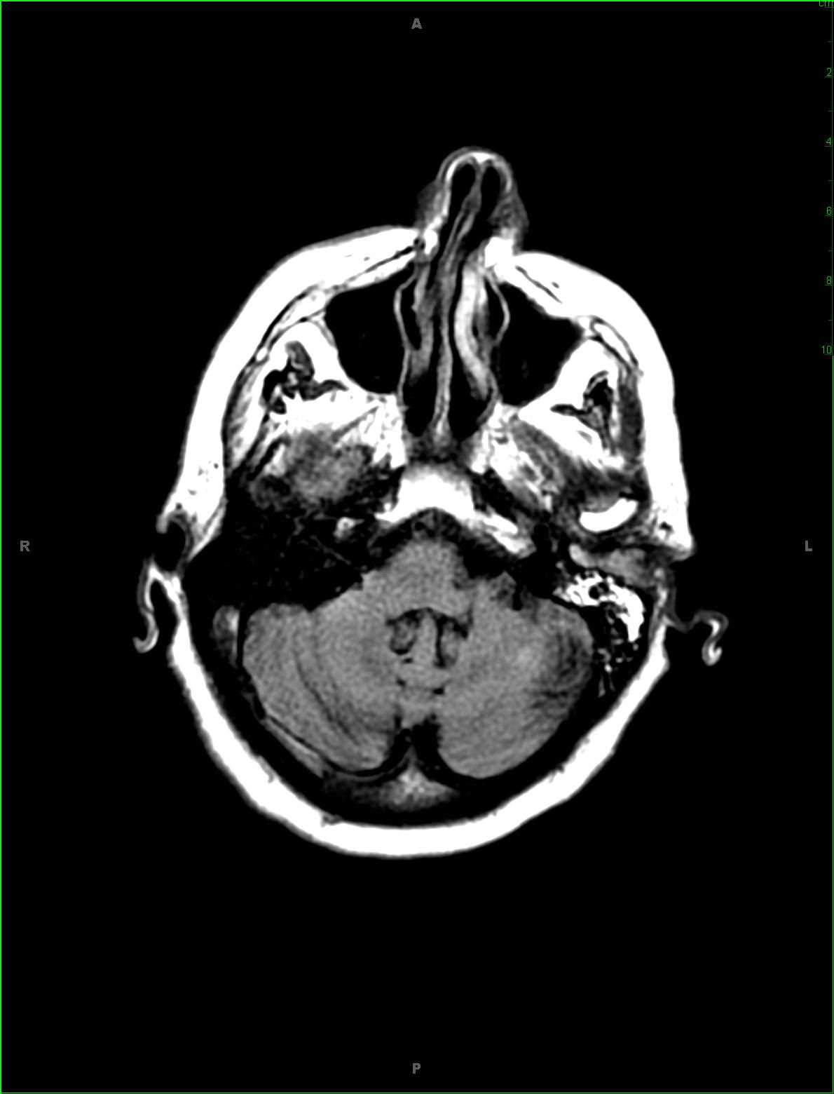
This is an unusual case of recurrent adenoid cystic carcinoma of the left external auditory canal in an 82-year-old male. The patient initially presented for gradual left-sided hearing loss. There is a slightly hyperintense soft tissue mass immediately posterior to the left mandibular condyle on the sagittal T1-weighted sequence, image 1, and non contrast fat-saturated T1-weighted sequence, image 2. Image 3, an axial FLAIR image, demonstrates an infiltrating slightly hyperintense lesion. There is no evidence of diffusion restriction on the ADC map, image 4. The lesion, which is predominantly involves the osseous portion of the external auditory canal with extension towards the cartilaginous segment, demonstrates mild postcontrast enhancement on images 5 and 6. Adenoid cystic carcinoma more commonly arises within the salivary glands, with the submandibular, sublingual, and minor salivary glands more commonly affected than the parotid gland. Perineural spread is common in adenoid cystic carcinoma, often resulting in disease recurrences after complete surgical resections.
Related videos to the case