- 2
- ,
- 2
- 4
- 6
To Quiz Yourself: Select OFF by clicking the button to hide the diagnosis & additional resources under the case.
Quick Browser: Select ON by clicking the button to hide the additional resources for faster case review.
CASE NUMBER
129


Diagnosis
Choroid Plexus Papilloma
Note
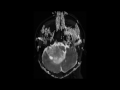
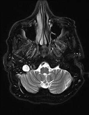
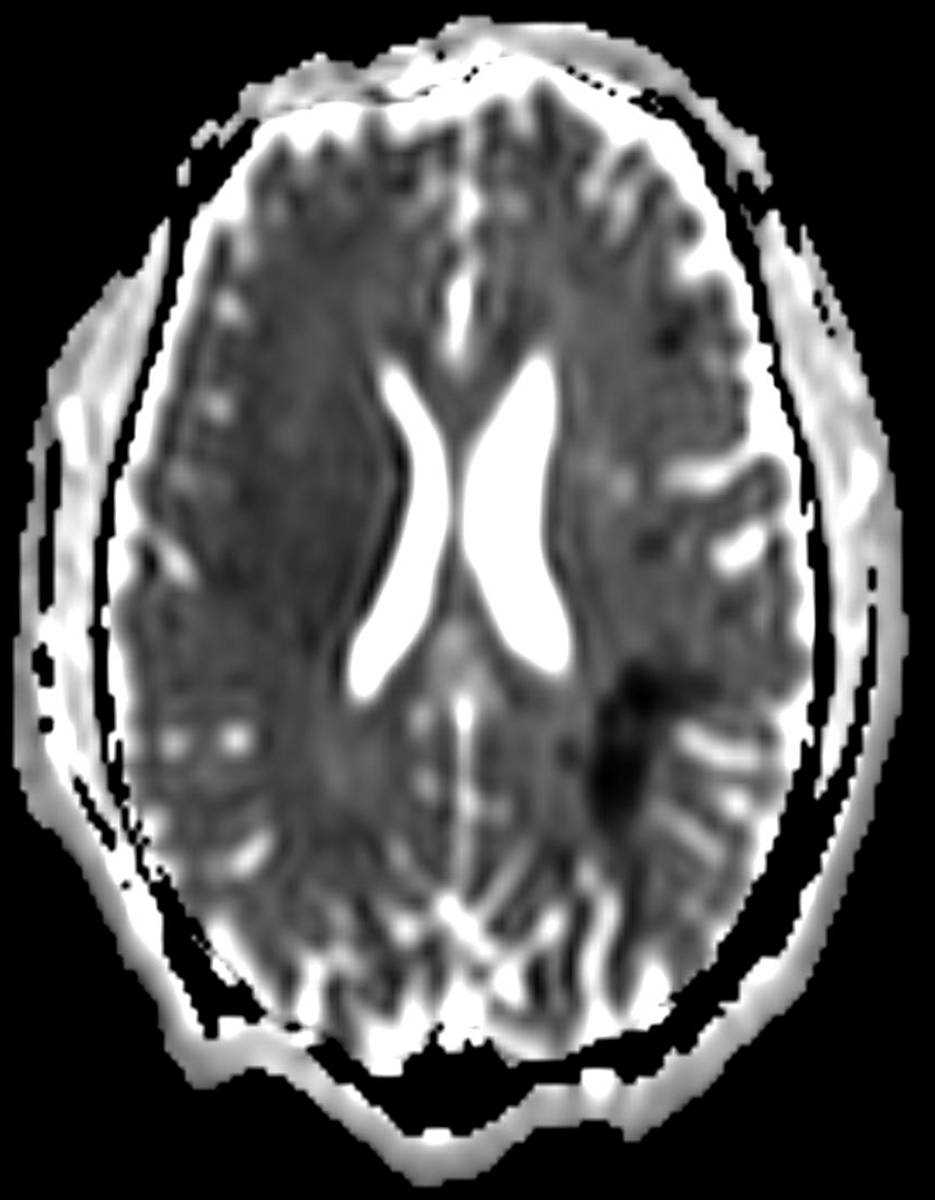
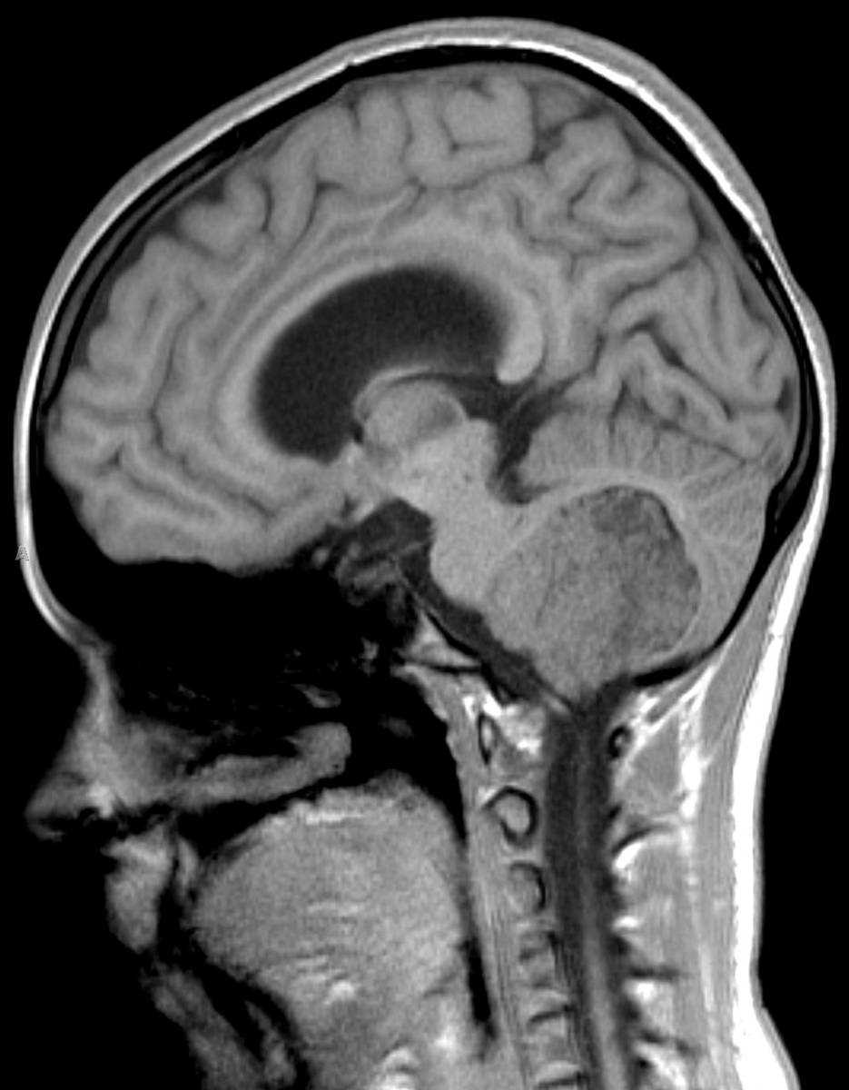
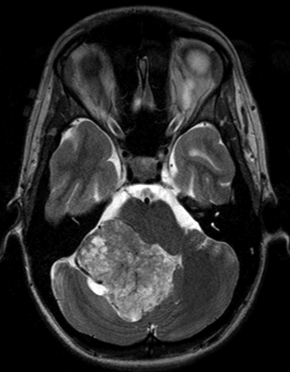
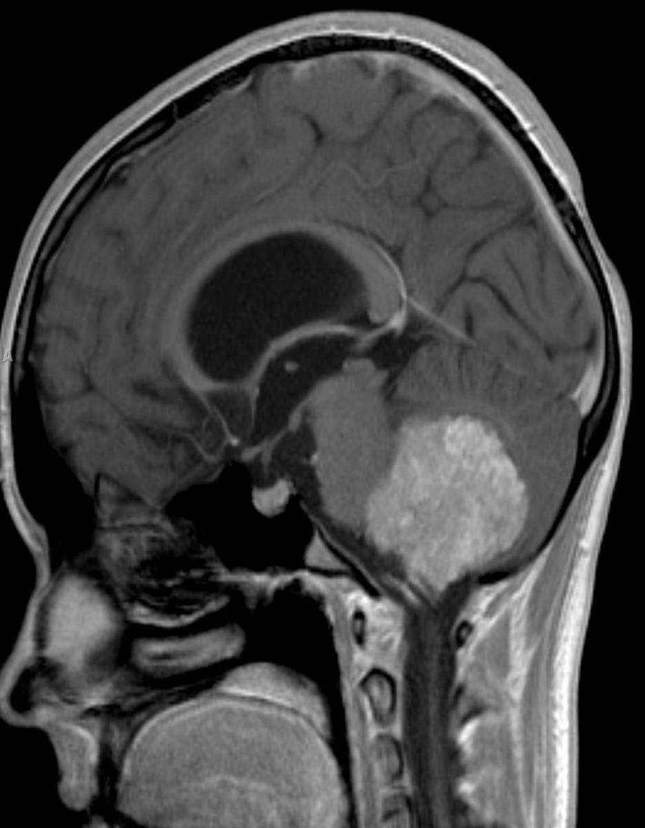
In this 3-year-old male with new onset symptoms of obstructive hydrocephalus, there is a circumscribed T1-isointense mass arising within, enlarging, and obliterating the fourth ventricle on the first image. Image 2, which is an axial FLAIR demonstrates a mildly hyperintense mass with several flow voids predominantly along the periphery of the lesion. The mass extends through and enlarges the foramen of Lushka on the right. There are no suspicious findings on the DWI or ADC maps, images 3 and 4, respectively. A small focal cystic region is identified along the posterolateral aspect of the lesion on the right. The presence of numerous flow voids, representing enlarged and tortuous feeding arteries, is confirmed on image 5, an axial T2-weighted sequence through the posterior cranial fossa. Image 6, a postcontrast sagittal T1-weighted sequence through the posterior cranial fossa demonstrates avid enhancement of the lesion. Choroid plexus papillomas, which are considered WHO grade I, make up about 3% of intracranial tumors in children. Greater than 80% of these lesions are identified in patients less than 5 years of age. The most common location choroid plexus papillomas are identified are at the trigone of the lateral ventricles. The lesions arise within the fourth ventricle more commonly in adults.
Related videos to the case