- 2
- ,
- 2
- 4
- 6
To Quiz Yourself: Select OFF by clicking the button to hide the diagnosis & additional resources under the case.
Quick Browser: Select ON by clicking the button to hide the additional resources for faster case review.
CASE NUMBER
127


Diagnosis
Intracranial Hypotension
Note
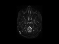
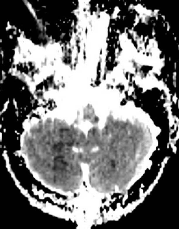
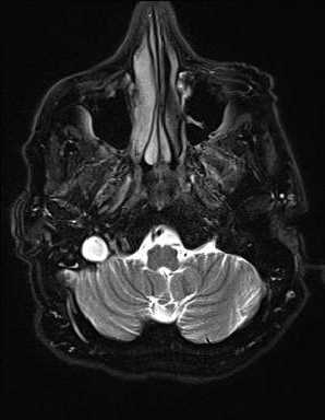
This is a case of intracranial hypotension in an 18-year-old female following ventriculoperitoneal shunt placement. The first image through the supratentorial compartment demonstrates a slit-like appearance of the frontal horns of the lateral ventricles. There is a general fullness and partial effacement of the cerebral cortical sulci. More inferiorly, on the second image through the posterior cranial fossa, we see that the overall effacement of the extra-axial CSF spaces continues, and the fourth ventricle is completely effaced. On the third image with axial T2 weighting through the level of the foramen magnum, there is crowding of the foramen with descent of the cerebellar tonsils. The fourth image, a sagittal CISS sequence, demonstrates sagging of the brainstem and fullness of the pituitary gland. Inferior descent of the supratentorial structures is confirmed on the coronal T1-weighted sequence, image 5, where there is mild downward transtentorial herniation of the uncus bilaterally. The final image, a postcontrast sagittal T1-weighted sequence, redemonstrates brainstem sagging, cerebellar tonsillar crowding, and increased pachymeningeal thickening and enhancement. Intracranial hypotension classically presents as a positional headache relieved with recumbent positioning. The opening pressure at LP will be decreased. Bilateral subdural hygromas may also be visualized.
Related videos to the case