- 2
- ,
- 2
- 4
- 6
To Quiz Yourself: Select OFF by clicking the button to hide the diagnosis & additional resources under the case.
Quick Browser: Select ON by clicking the button to hide the additional resources for faster case review.
CASE NUMBER
128


Diagnosis
Transverse/Sigmoid Sinus Venous Thrombosis
Note
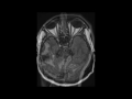
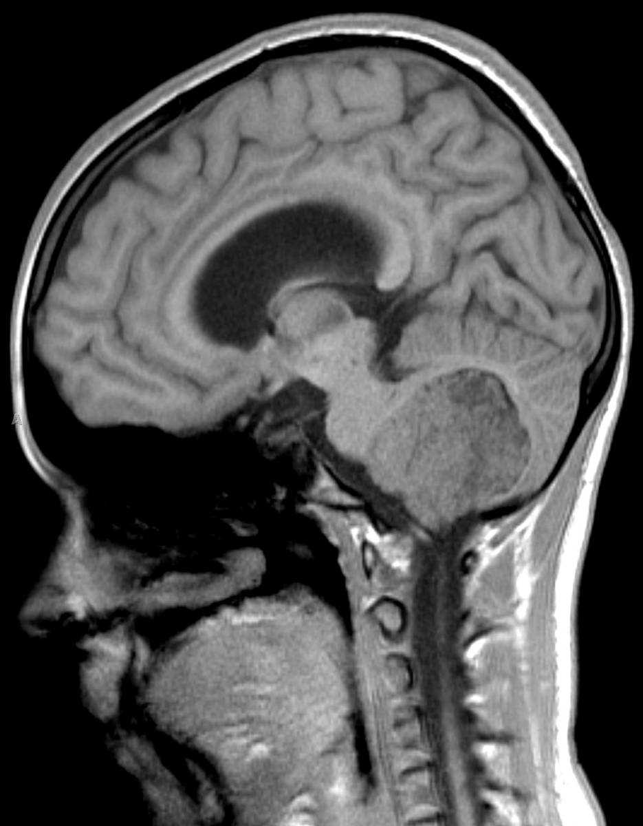
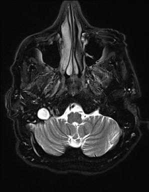
This is a case of right-sided transverse, sigmoid, and jugular venous thrombosis presenting in a 66-year-old male with a history of pancreatic neuroendocrine neoplasm. On the axial T2-weighted image, there is increased T2 signal intensity in the distal transverse and proximal right sigmoid sinus on the right with additional increased T2 signal intensity at the proximal right internal jugular bulb. As we can see on the left, the normal appearance of the dural venous sinuses is hypointense on the T2-weighted sequences, due to the presence of venous flow. The second image, with FLAIR weighting, demonstrates increased FLAIR signal hyperintensity in the right transverse sinus. The third image, which is sagittal T1-weighted and off midline, demonstrates hyperintense signal compatible with thrombus extending from the right jugular bulb more inferiorly. The findings are confirmed on the DWI and ADC map, images four and five, where there is diffusion restriction at the sites of thrombosis. The 3D maxim intensity projection MR venogram confirms absence of flow within the thrombosed transverse sinus, sigmoid sinus, and internal jugular vein on the right. Causes of venous sinus thrombosis include: acute dehydration, chemotherapeutic agents, hypercoagulable states, iatrogenic causes, infection, malignancy as in our case, malnutrition, pregnancy, and trauma, among others.
Related videos to the case