- 2
- ,
- 2
- 4
- 6
To Quiz Yourself: Select OFF by clicking the button to hide the diagnosis & additional resources under the case.
Quick Browser: Select ON by clicking the button to hide the additional resources for faster case review.
CASE NUMBER
361


Diagnosis
Intracranial Vasospasm
Note
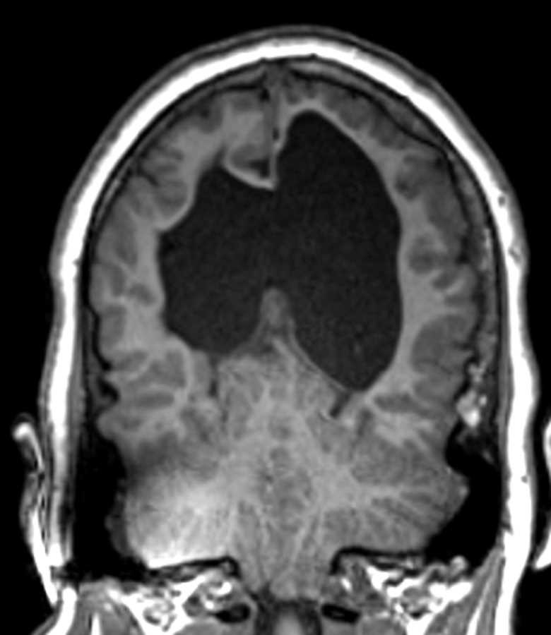
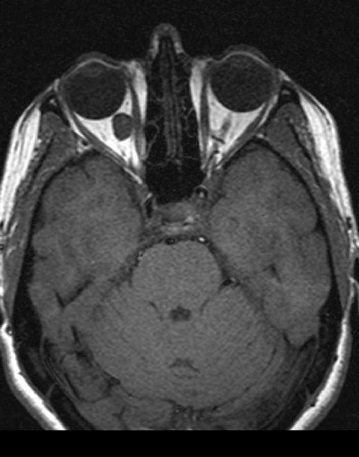
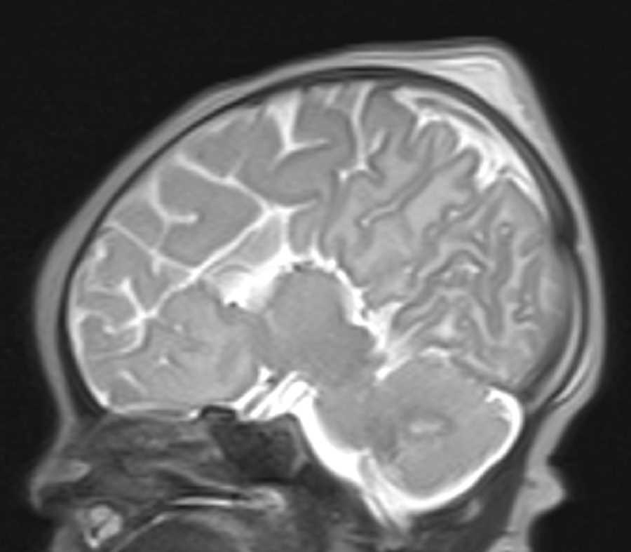
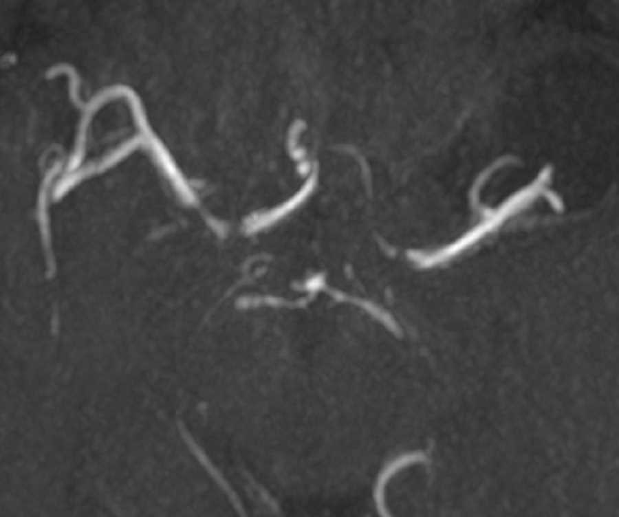
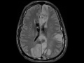
These images demonstrate diffuse subarachnoid hemorrhage, large intraventricular hemorrhage, and small bilateral subdural hemorrhages as evidenced by decreased signal on the susceptibility weighted images. There are multifocal areas of restricted diffusion and associated mildly expansile T2 FLAIR hyperintensity throughout the supratentorial brain, cerebellum, and brainstem. Axial 3D time of flight imaging of the circle of willis with multiplanar maximum intensity projection images shows multifocal areas of narrowing involving the bilateral internal carotid artery termini, proximal M1 segments, proximal A2 segments, distal right V4 segment, and right P2 segment. Findings are compatible with vasospasm and extensive associated ischemic infarcts secondary to diffuse subarachnoid hemorrhage from a ruptured aneurysm found in the left V4 segment which was status post coiling.
Related videos to the case

THIS IS CASE
361
OF
373