- 2
- ,
- 2
- 4
- 6
To Quiz Yourself: Select OFF by clicking the button to hide the diagnosis & additional resources under the case.
Quick Browser: Select ON by clicking the button to hide the additional resources for faster case review.
CASE NUMBER
343


Diagnosis
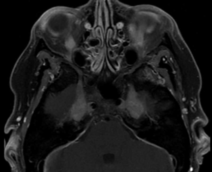
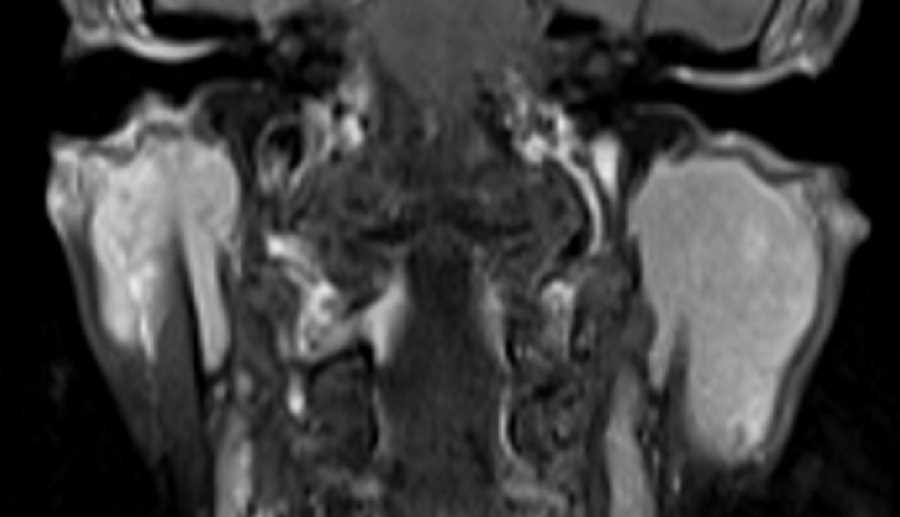
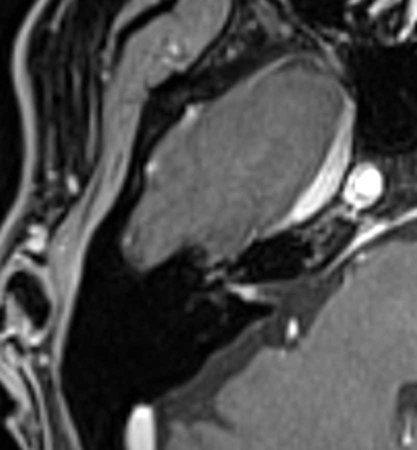
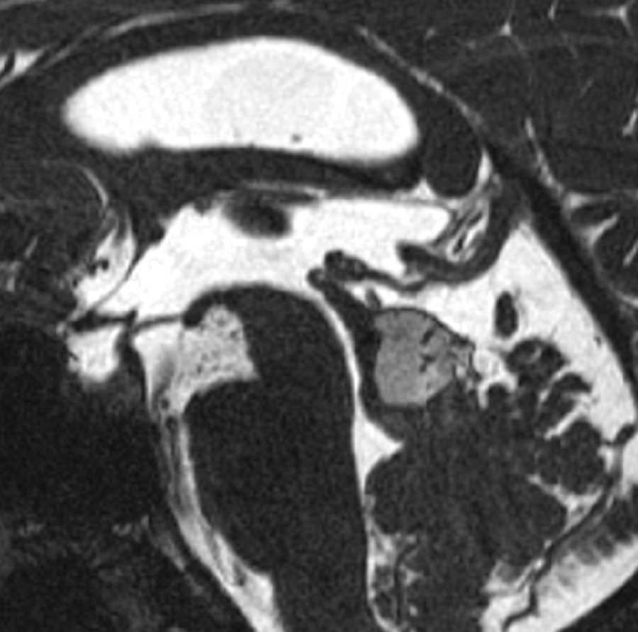
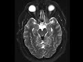
Tectal Plate Lipoma
Note
These images show a T1 hypertense, T2 FLAIR hyperintense mass of the tectal plate which becomes hypointense on T2 fat-saturated images compatible with a lipoma. High resolution sagittal CISS imaging shows the cerebral aqueduct to be patent. About half of intracranial lipomas are associated with the corpus callosum with suprasellar and tectal locations making up the second and third most common locations. These masses are usually found incidentally and the tubulonodular type in the pericallosal location may demonstrate rim calcification and be seen with anomalies of the corpus callosum.
Related videos to the case

THIS IS CASE
343
OF
373