- 2
- ,
- 2
- 4
- 6
To Quiz Yourself: Select OFF by clicking the button to hide the diagnosis & additional resources under the case.
Quick Browser: Select ON by clicking the button to hide the additional resources for faster case review.
CASE NUMBER
337


Diagnosis
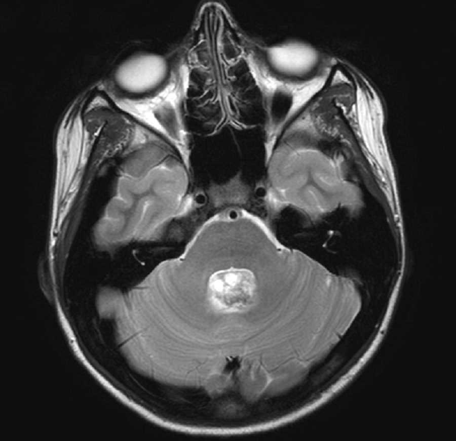
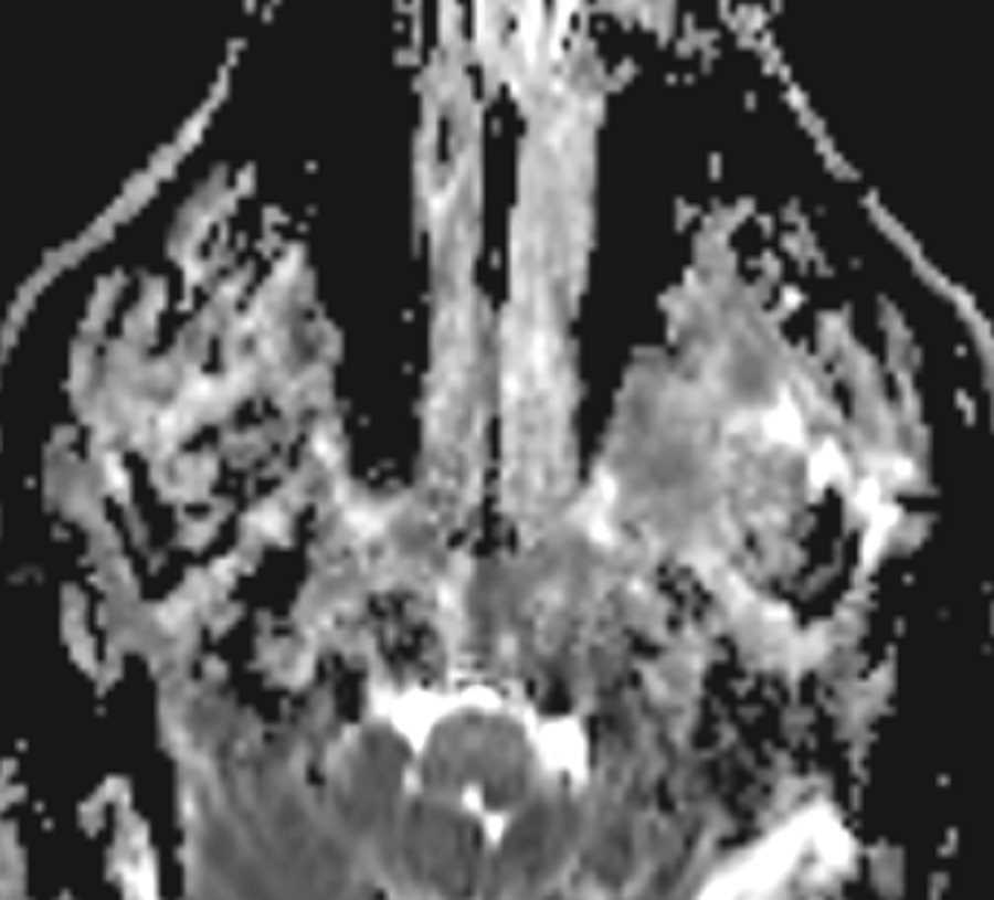
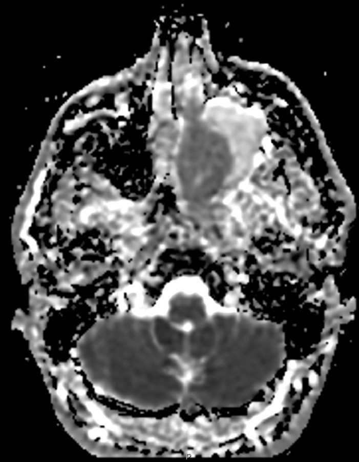
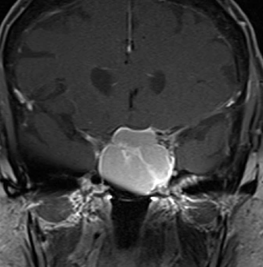
Sphenoid Mucocele
Note
These images demonstrate a large T1 hyperintense, T2 hypointense mass which expands the sphenoid sinus distorting and lifting the anterior sella and adjacent skull base as well as the inferomedial left orbital wall. Post contrast images shows characteristic peripheral mucosal enhancement characteristic of mucocele. The T1 and T2 characteristics show that the mucocele contents are proteinaceous. The differential considerations include a large sellar mass protruding into the sphenoid sinus such as a adenoma, a clival mass, or sinonasal neoplasm. The sphenoid location for mucocele is the least common as most occur in the frontal sinuses.
Related videos to the case


THIS IS CASE
337
OF
373