- 2
- ,
- 3
- 8
- 1
To Quiz Yourself: Select OFF by clicking the button to hide the diagnosis & additional resources under the case.
Quick Browser: Select ON by clicking the button to hide the additional resources for faster case review.
CASE NUMBER
351


Diagnosis
Carotid Cavernous Fistula
Note
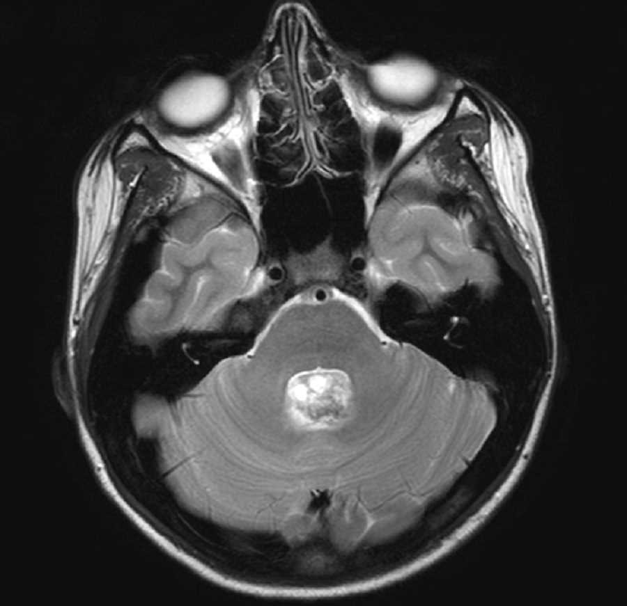
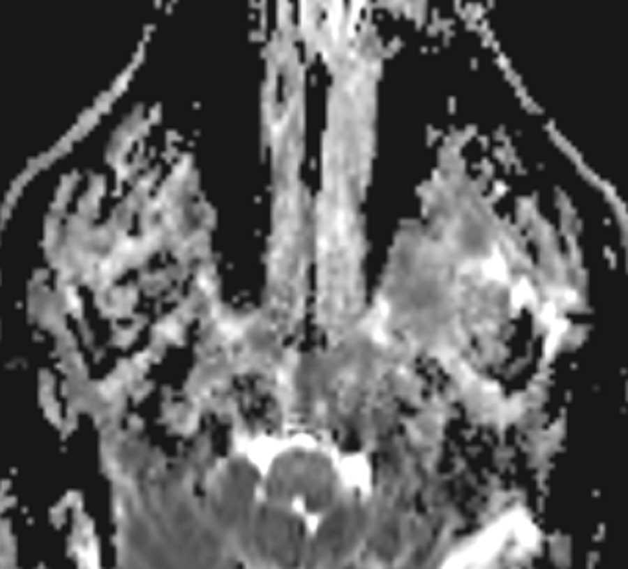
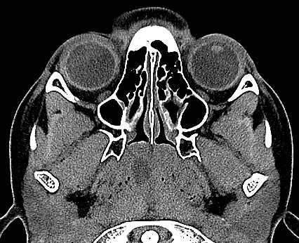
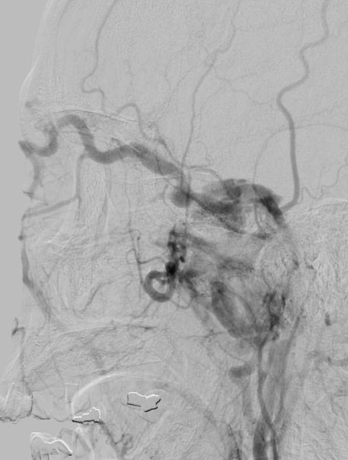
These first five CTA images of the head demonstrate filling of the left cavernous sinus in the arterial phase and asymmetric enlargement and filling of the left superior ophthalmic vein. These findings are consistent with a carotid cavernous fistula. The diagnostic angiogram was performed to evaluate the supply and drainage of the fistula. No aneurysm was identified. The fistula was supplied most prominently from the bilateral external carotid arteries and showed prominent retrograde drainage into the dilated left superior ophthalmic vein. These fistulas may present with unilateral or bilateral proptosis and chemosis and if severe may cause vision loss. This patient was treated with transvenous coil embolization and demonstrated no evidence of fistula on two month follow-up imaging.
Related videos to the case


THIS IS CASE
351
OF
396