- 2
- ,
- 3
- 8
- 1
To Quiz Yourself: Select OFF by clicking the button to hide the diagnosis & additional resources under the case.
Quick Browser: Select ON by clicking the button to hide the additional resources for faster case review.
CASE NUMBER
348


Diagnosis
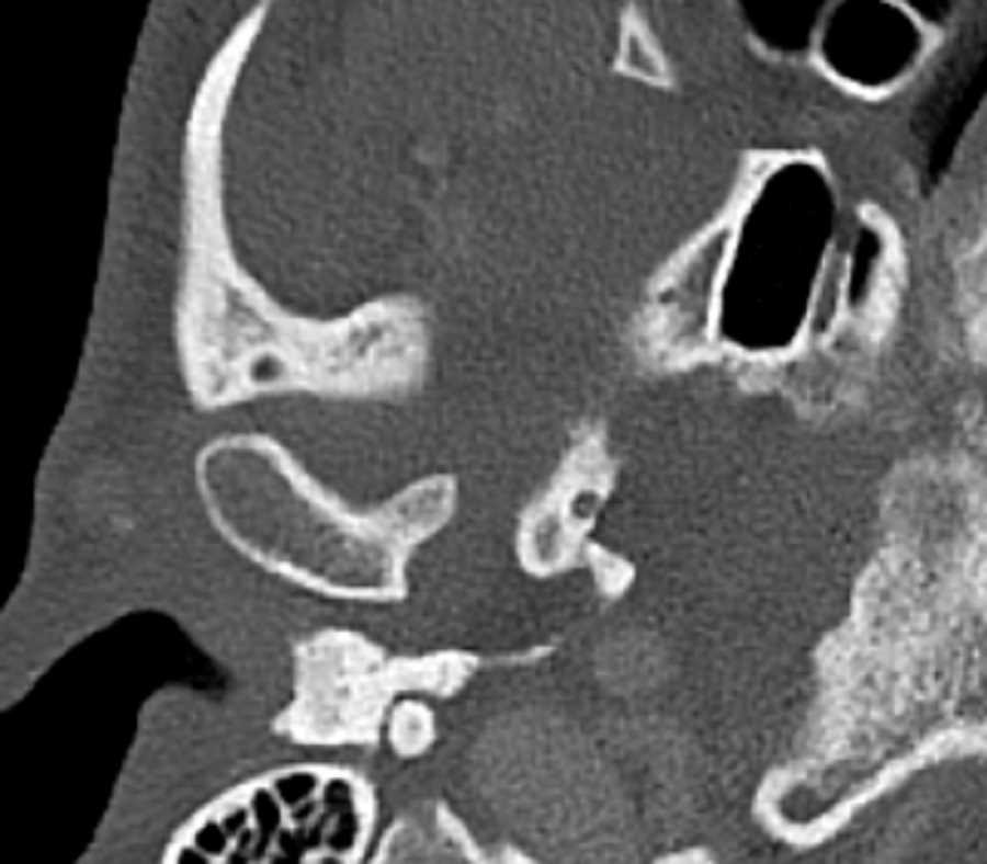
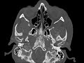
Mandibular Condyle Osteochondroma
Note
These images demonstrate a small non-articulating exostosis emanating from the anteromedial aspect of the right mandibular condyle. The cortex and medullary bone is contiguous with that of the mandibular condyle. Findings are most compatible with a small osteochondroma. The coronoid process and condyle are the most common sites for lesions affecting the jaw. The differential would include osteophyte in degenerative joint disease, condylar hyperplasia, and osteoma. Larger lesions may result in facial asymmetry and abnormal bite.
Related videos to the case

THIS IS CASE
348
OF
396