- 2
- ,
- 2
- 5
- 2
To Quiz Yourself: Select OFF by clicking the button to hide the diagnosis & additional resources under the case.
Quick Browser: Select ON by clicking the button to hide the additional resources for faster case review.
CASE NUMBER
334


Diagnosis
Venous Malformations
Note
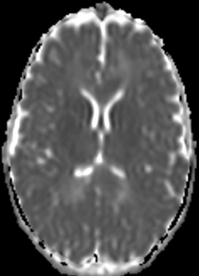
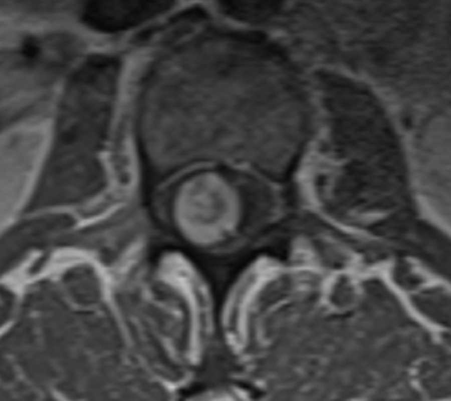
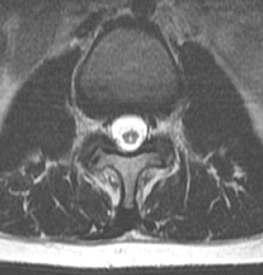
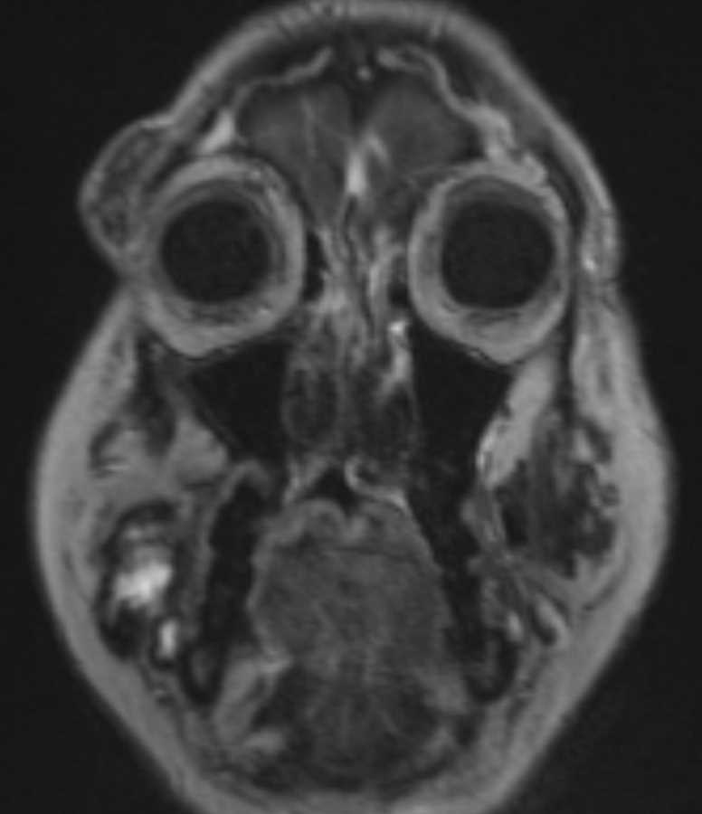
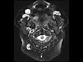
These images show multiple T2 hyperintense extracranial lesions involving the right periorbital soft tissues, right buccal region abutting the right facial vein, and in the left masticator space anterior to the masseter. The lesion in the right periorbital soft tissues shows a fluid level and minimal nodular venous enhancement inferiorly. The lesion anterior to the left masseter muscle shows two rounded signal voids corresponding to calcifications see on a CT not pictured here which are compatible with phleboliths. Time of flight imaging shows no evidence of arterial supply to these lesions. Findings are most compatible with multiple venous malformations with phleboliths being a characteristic feature. Venous malformations of the head and neck may be transpatial or localized and are most commonly found in the buccal region as seen on the right in this patient with additional common sites including the submandibular and masticator spaces.
Related videos to the case

THIS IS CASE
334
OF
374