
- 3
- 2
- 6
- ,
- 4
- 8
- 9
Explore our numerous CT Scans, MRI, and X-Rays with the diagnosis in different anatomical regions and other Body CT topics. These case studies are designed for the education of medical professionals and radiologist.

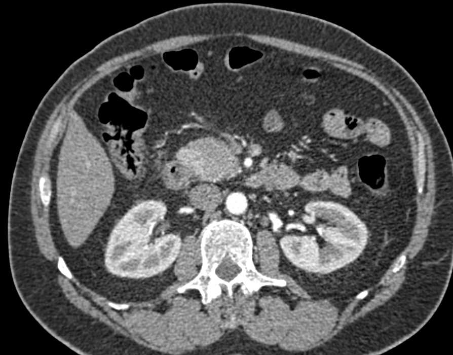
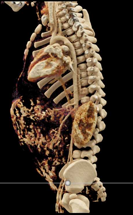
Adrenal
+ 7,418 Teaching File Images
+ 11,081,369 Views
+ Most Recent Case: Epithelioid Angiosarcoma
+ 7,418 Teaching File Images
+ 11,081,369 Views
+ Most Recent Case: Epithelioid Angiosarcoma
Body MR
+ 2,321 Teaching File Images
+ 4,675,691 Views
+ Most Recent Case: Carcinoma of the Head of the ...
+ 2,321 Teaching File Images
+ 4,675,691 Views
+ Most Recent Case: Carcinoma of the Head of the ...

Cardiac
+ 24,607 Teaching File Images
+ 30,615,427 Views
+ Most Recent Case: Cinematic Rendering of Normal...
+ 24,607 Teaching File Images
+ 30,615,427 Views
+ Most Recent Case: Cinematic Rendering of Normal...

Chest
+ 30,679 Teaching File Images
+ 43,776,067 Views
+ Most Recent Case: Enlarged Bronchial Arteries
+ 30,679 Teaching File Images
+ 43,776,067 Views
+ Most Recent Case: Enlarged Bronchial Arteries

Colon
+ 10,677 Teaching File Images
+ 16,984,389 Views
+ Most Recent Case: Rectal Bleed From Varices
+ 10,677 Teaching File Images
+ 16,984,389 Views
+ Most Recent Case: Rectal Bleed From Varices


Gastrointestinal
+ 11,691 Teaching File Images
+ 15,197,934 Views
+ Most Recent Case: Pulmonary Embolism in Post Op...
+ 11,691 Teaching File Images
+ 15,197,934 Views
+ Most Recent Case: Pulmonary Embolism in Post Op...

Genitourinary
+ 13,567 Teaching File Images
+ 15,878,108 Views
+ Most Recent Case: Penile Prosthesis With CR
+ 13,567 Teaching File Images
+ 15,878,108 Views
+ Most Recent Case: Penile Prosthesis With CR

Kidney
+ 51,591 Teaching File Images
+ 58,181,532 Views
+ Most Recent Case: Parapelvic Cysts in the Kidne...
+ 51,591 Teaching File Images
+ 58,181,532 Views
+ Most Recent Case: Parapelvic Cysts in the Kidne...

Liver
+ 33,702 Teaching File Images
+ 44,234,393 Views
+ Most Recent Case: Vascular Metastases to the Li...
+ 33,702 Teaching File Images
+ 44,234,393 Views
+ Most Recent Case: Vascular Metastases to the Li...

Musculoskeletal
+ 35,870 Teaching File Images
+ 41,880,433 Views
+ Most Recent Case: Rectus Sheath Hematoma in A P...
+ 35,870 Teaching File Images
+ 41,880,433 Views
+ Most Recent Case: Rectus Sheath Hematoma in A P...


Neuro MR
+ 2,252 Teaching File Images
+ 3,880,754 Views
+ Most Recent Case: Pituitary Adenoma Secretes GH...
+ 2,252 Teaching File Images
+ 3,880,754 Views
+ Most Recent Case: Pituitary Adenoma Secretes GH...
OB/GYN
+ 2,827 Teaching File Images
+ 4,920,919 Views
+ Most Recent Case: Rectus Sheath Hematoma in A P...
+ 2,827 Teaching File Images
+ 4,920,919 Views
+ Most Recent Case: Rectus Sheath Hematoma in A P...

Pancreas
+ 47,037 Teaching File Images
+ 37,989,963 Views
+ Most Recent Case: Carcinoma Body of Pancreas Wi...
+ 47,037 Teaching File Images
+ 37,989,963 Views
+ Most Recent Case: Carcinoma Body of Pancreas Wi...
Pediatric
+ 480 Teaching File Images
+ 1,583,492 Views
+ Most Recent Case: Ductus Bump But Patent Ductus...
+ 480 Teaching File Images
+ 1,583,492 Views
+ Most Recent Case: Ductus Bump But Patent Ductus...

Spleen
+ 8,837 Teaching File Images
+ 13,837,190 Views
+ Most Recent Case: Vascular Metastases to the Li...
+ 8,837 Teaching File Images
+ 13,837,190 Views
+ Most Recent Case: Vascular Metastases to the Li...

Stomach
+ 11,738 Teaching File Images
+ 14,889,575 Views
+ Most Recent Case: Gastric Cancer With Perforati...
+ 11,738 Teaching File Images
+ 14,889,575 Views
+ Most Recent Case: Gastric Cancer With Perforati...

Vascular
+ 68,006 Teaching File Images
+ 68,200,685 Views
+ Most Recent Case: Enlarged Bronchial Arteries
+ 68,006 Teaching File Images
+ 68,200,685 Views
+ Most Recent Case: Enlarged Bronchial Arteries

Veterinary
+ 2,338 Teaching File Images
+ 3,116,112 Views
+ Most Recent Case: CT Turtle With Nice Musculosk...
+ 2,338 Teaching File Images
+ 3,116,112 Views
+ Most Recent Case: CT Turtle With Nice Musculosk...
Virtual Imaging
+ 218 Teaching File Images
+ 1,854,721 Views
+ Most Recent Case: Virtual Colon: Large 1.6 Cm P...
+ 218 Teaching File Images
+ 1,854,721 Views
+ Most Recent Case: Virtual Colon: Large 1.6 Cm P...
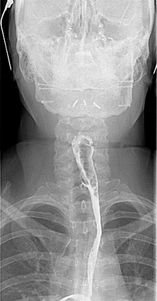
X Rays
+ 316 Teaching File Images
+ 2,142,733 Views
+ Most Recent Case: Perforated Esophagus Due to F...
+ 316 Teaching File Images
+ 2,142,733 Views
+ Most Recent Case: Perforated Esophagus Due to F...