- 2
- ,
- 2
- 5
- 2
To Quiz Yourself: Select OFF by clicking the button to hide the diagnosis & additional resources under the case.
Quick Browser: Select ON by clicking the button to hide the additional resources for faster case review.
CASE NUMBER
325


Diagnosis
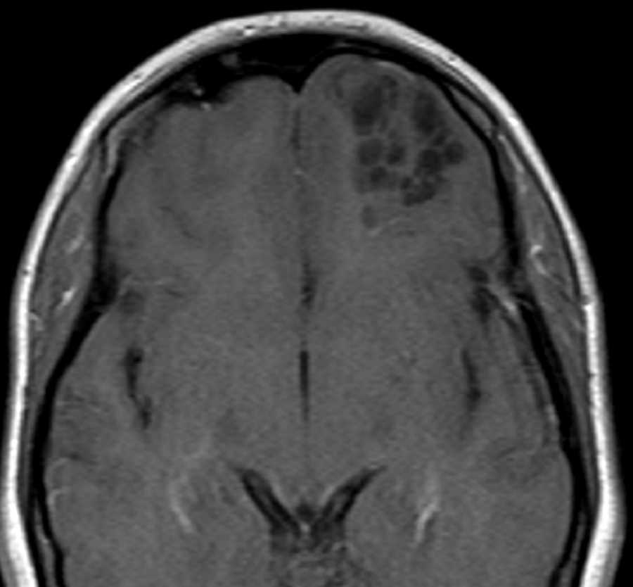
Dysembryoplastic neuroepithelial tumor (DNET)
Note
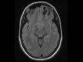
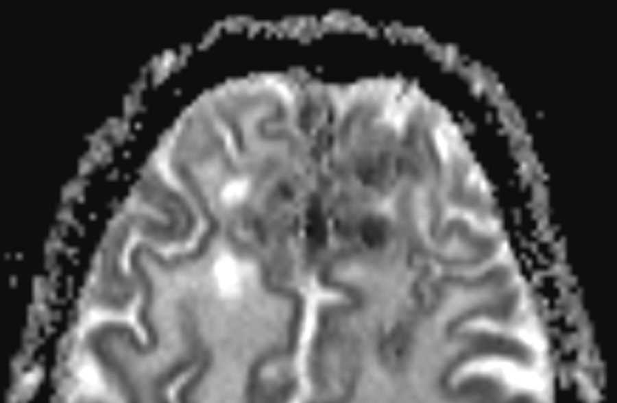
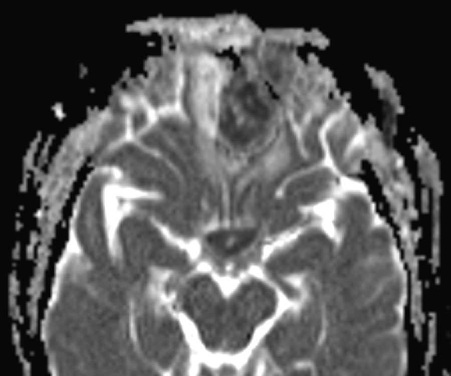
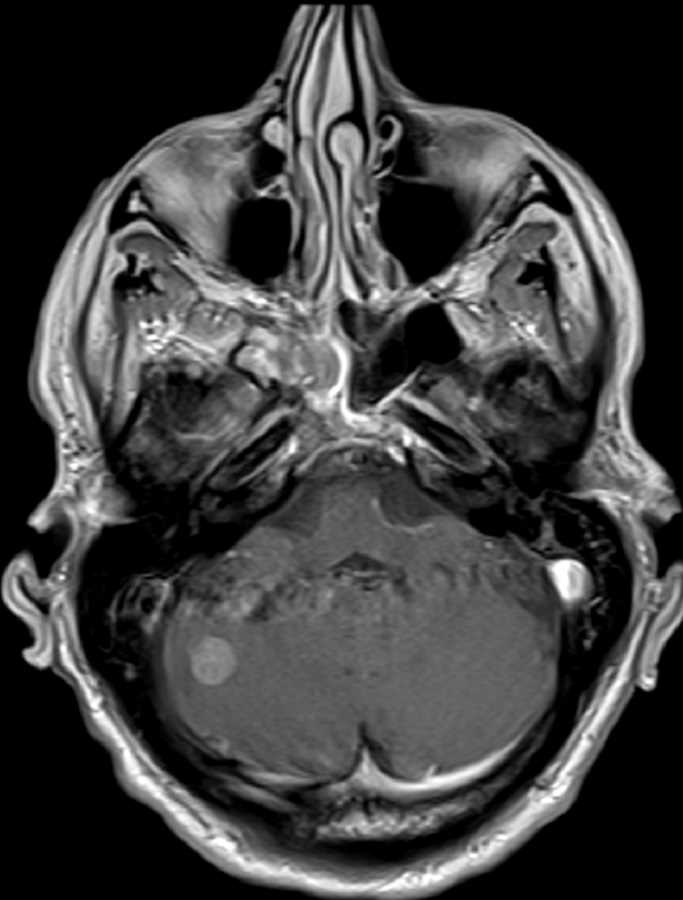
These images show a wedge-shaped, multi-cystic, expansile, non-enhancing, T2 hyperintense lesion involving the cortex and subcortical white matter of the anterior left frontal lobe. There is minimal surrounding T2 FLAIR hyperintensity which is most prominent deep to the lesion in the adjacent periventricular white matter. No restricted diffusion is present. In this young adult, this imaging appearance is most consistent with a benign entity such as dysembryoplastic neuroepithelial tumor (DNET) or giant tumefactive perivascular spaces. DNETs are usually wedge-shaped bubbly, cystic lesions pointing towards the ventricle, and usually do not demonstrate any enhancement. Most commonly they occur in the mesial temporal lobes, but may be found involving any supratentorial cortex. In patients who present with seizures, surgical resection can be performed and is usually curative.
Related videos to the case