- 2
- ,
- 3
- 8
- 1
To Quiz Yourself: Select OFF by clicking the button to hide the diagnosis & additional resources under the case.
Quick Browser: Select ON by clicking the button to hide the additional resources for faster case review.
CASE NUMBER
328
Diagnosis
Fibrous Dysplasia-MRI
Note
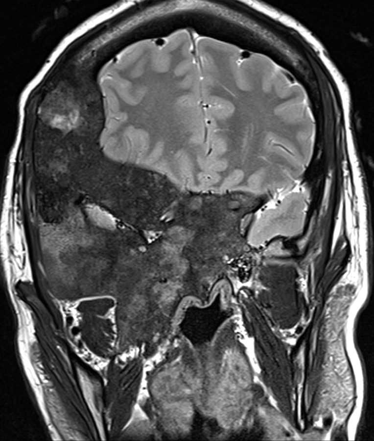
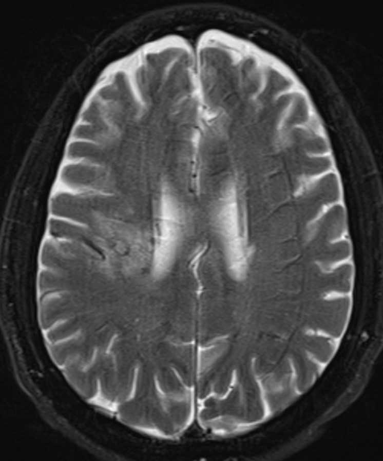
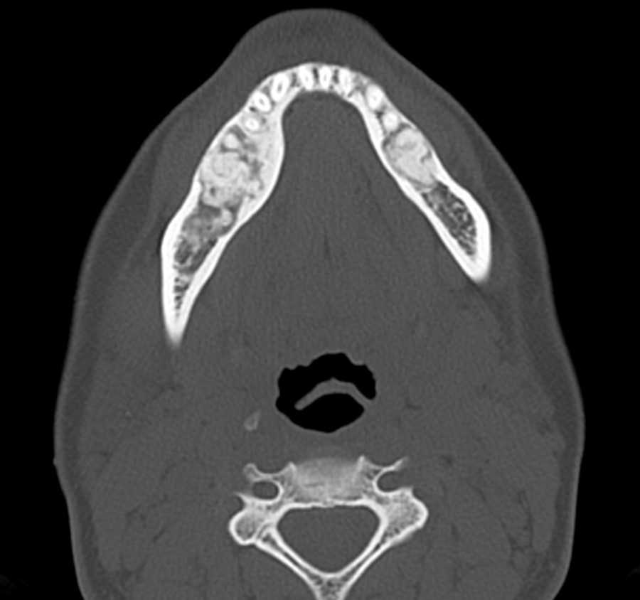
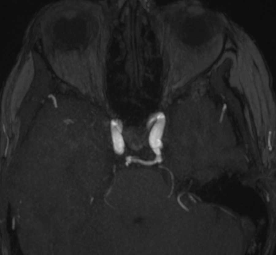
These images show a large extensive heterogeneously enhancing expansile osseous lesion which crosses sutures and involves the right frontal and temporal calvarium, bony orbit, right ethmoid, and maxillary sinuses resulting in severe facial deformity and marked right proptosis. The involved sinuses and mastoid air cells are obliterated. This heterogeneous appearance and enhancement on MRI can be potentially misleading to think of more aggressive neoplasms. Many times the CT appearance of ground-glass opacity helps to cinch the diagnosis of craniofacial fibrous dysplasia. The patient may develop symptoms related to cranial nerve palsies from narrowing of the skull base foramina.
THIS IS CASE
328
OF
396


