- 2
- ,
- 3
- 8
- 1
To Quiz Yourself: Select OFF by clicking the button to hide the diagnosis & additional resources under the case.
Quick Browser: Select ON by clicking the button to hide the additional resources for faster case review.
CASE NUMBER
322
Diagnosis
Sturge-Weber Syndrome
Note
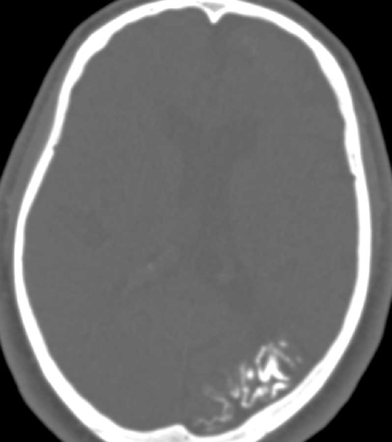
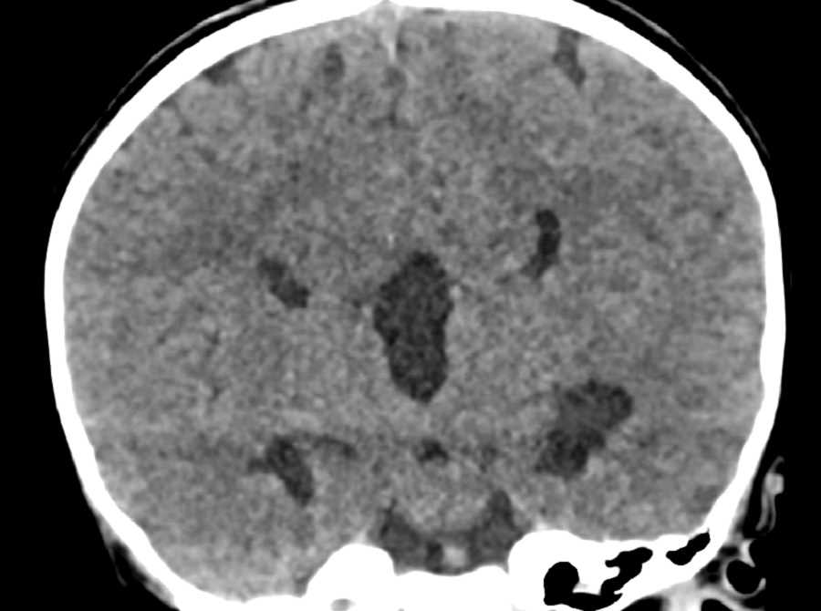
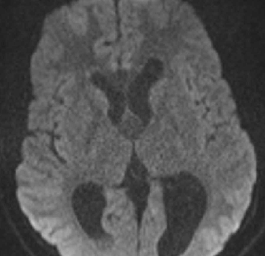
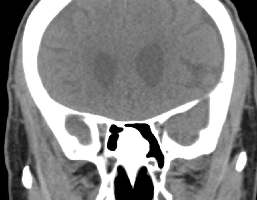
These images show cortical calcifications and atrophy of the left parietal and occipital lobes. This is a characteristic feature of Sturge-Weber Syndrome. This calcification results from development of a pial angioma that is generally parieto-occipital. Cortical veins fail to develop and stasis of blood results in dystrophic calcification leading to atrophy. This syndrome involves the face, brain, and meninges. Patients usually develop seizures.
THIS IS CASE
322
OF
396


