- 2
- ,
- 2
- 5
- 2
To Quiz Yourself: Select OFF by clicking the button to hide the diagnosis & additional resources under the case.
Quick Browser: Select ON by clicking the button to hide the additional resources for faster case review.
CASE NUMBER
283


Diagnosis
Granulomatous Inflammation, IgG4 Disease
Note
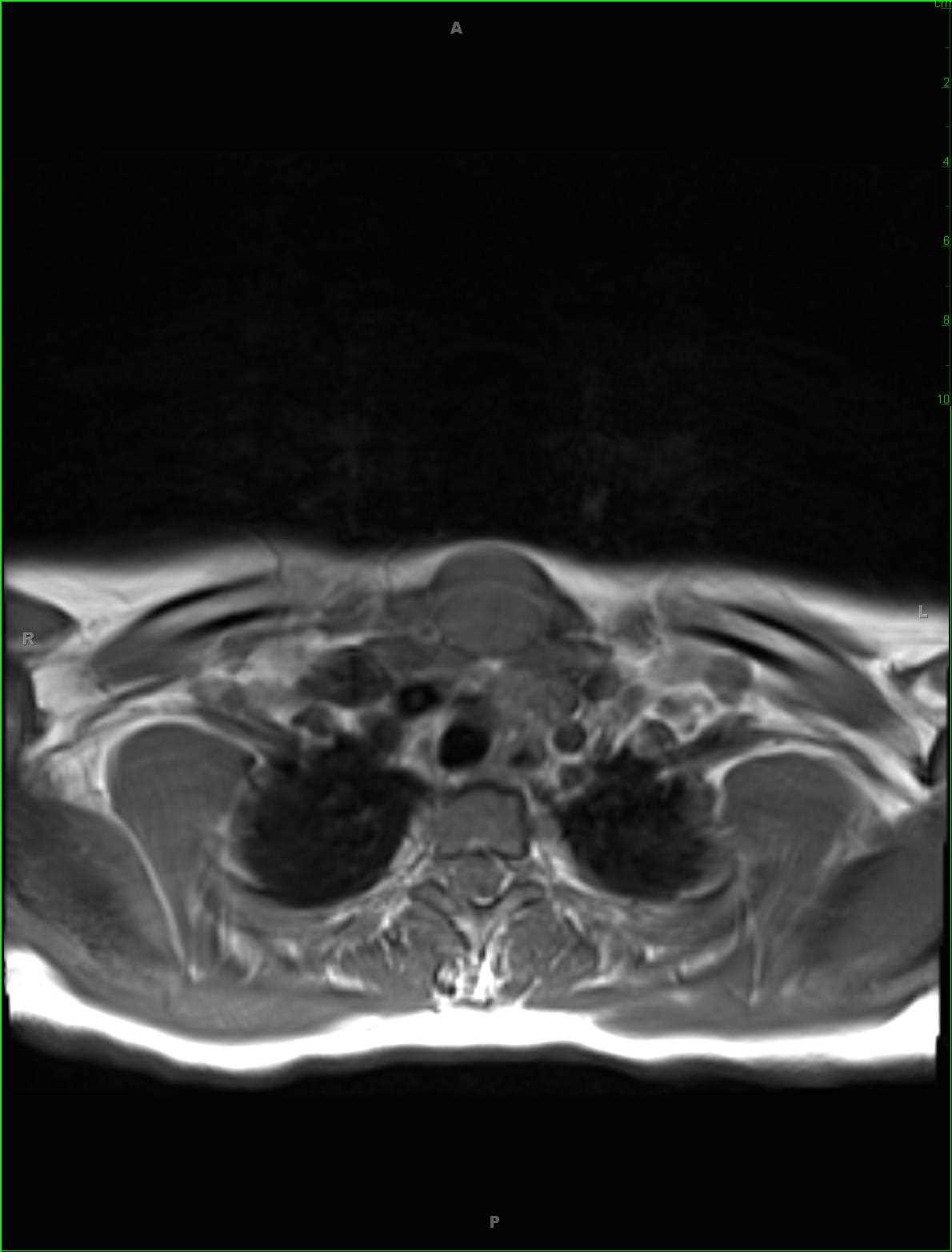
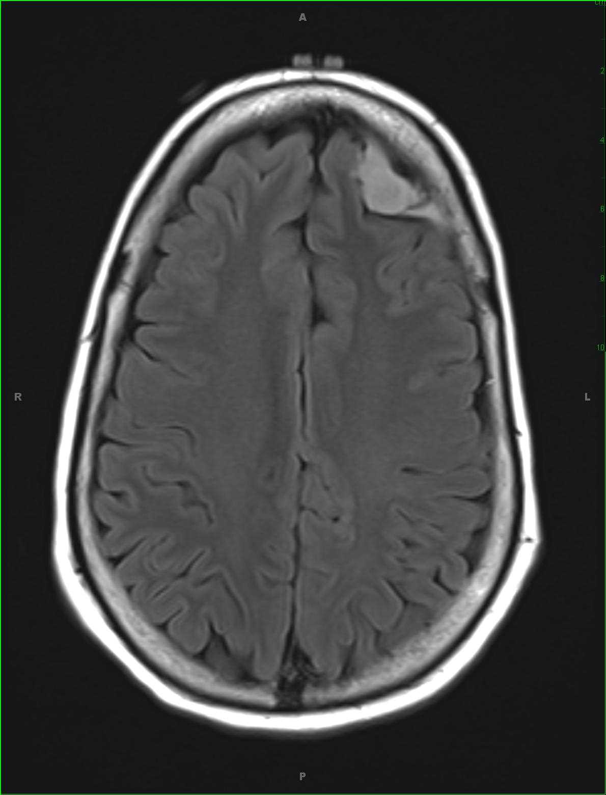
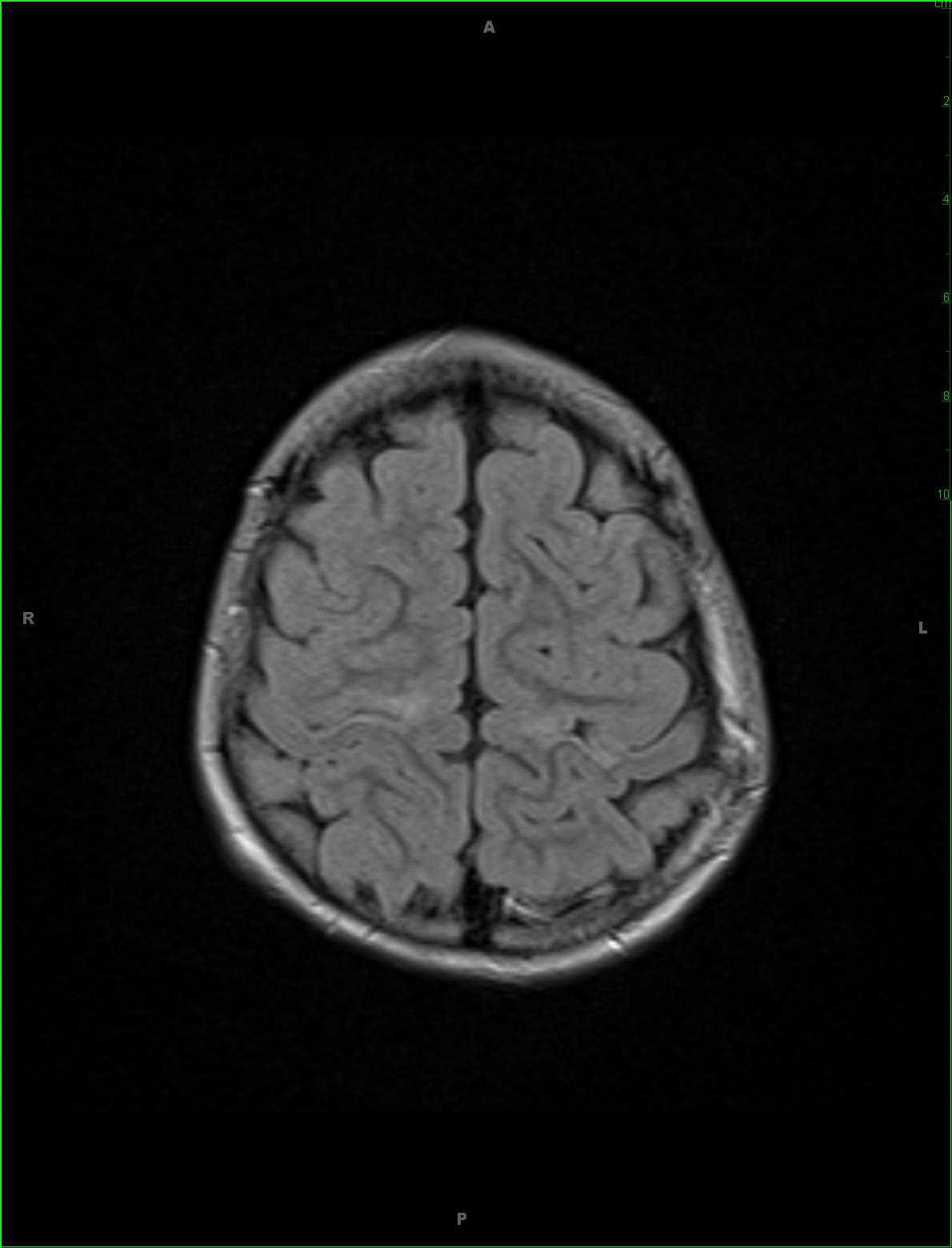
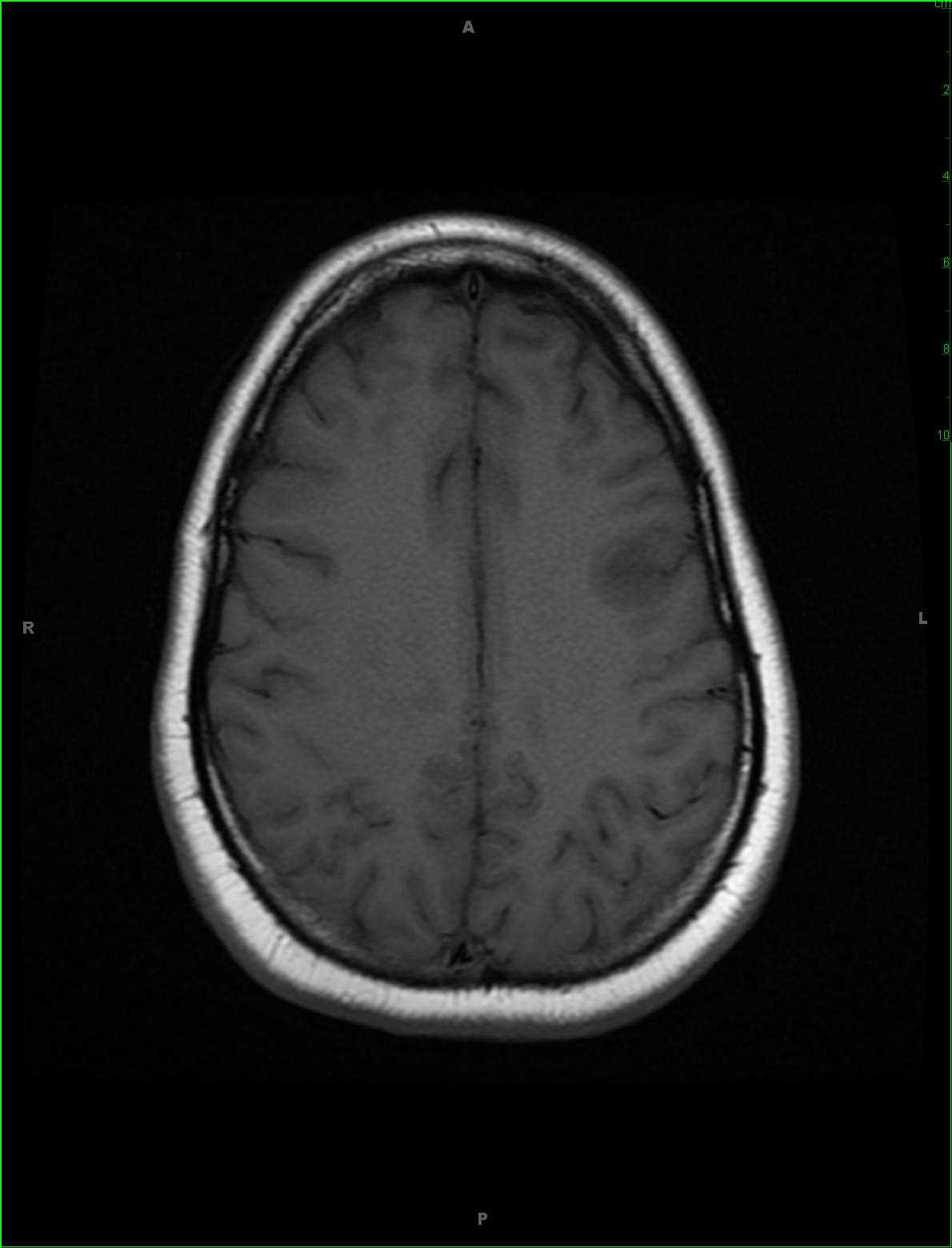
42-year-old female that presented with new onset seizures. There is a rounded T1 hypointense region centered within the cortical gray matter of the left middle frontal gyrus with extension to involve the juxtacortical white matter. The lesion is T2/FLAIR hyperintense and there is no signal loss on the susceptibility weighted images. No diffusion restriction. There is a confluent region of enhancement with serpiginous borders on the postcontrast sagittal image. A differential of oligodendroglioma, demyelinating plaque, and less likely metastatic disease was given. On biopsy, this lesion was found to be granulomatous inflammation in the setting of IgG4 disease. Immunoglobulin 4 related systemic disease is characterized by abundant infiltration of IgG4 positive plasma cells and lymphocytes with associated fibrosis. Head-neck manifestations include salivary gland, lacrimal gland, orbital, thyroid gland, lymph node, sinonasal cavity and pituitary stalk involvement. IgG 4 related disease in either the pituitary or brain parenchyma itself is rare. Treatment is with systemic corticosteroid therapy.
Related videos to the case