- 2
- ,
- 2
- 5
- 2
To Quiz Yourself: Select OFF by clicking the button to hide the diagnosis & additional resources under the case.
Quick Browser: Select ON by clicking the button to hide the additional resources for faster case review.
CASE NUMBER
284


Diagnosis
Prior Hypoxic Ischemic Insult with Wallerian Degeneration
Note
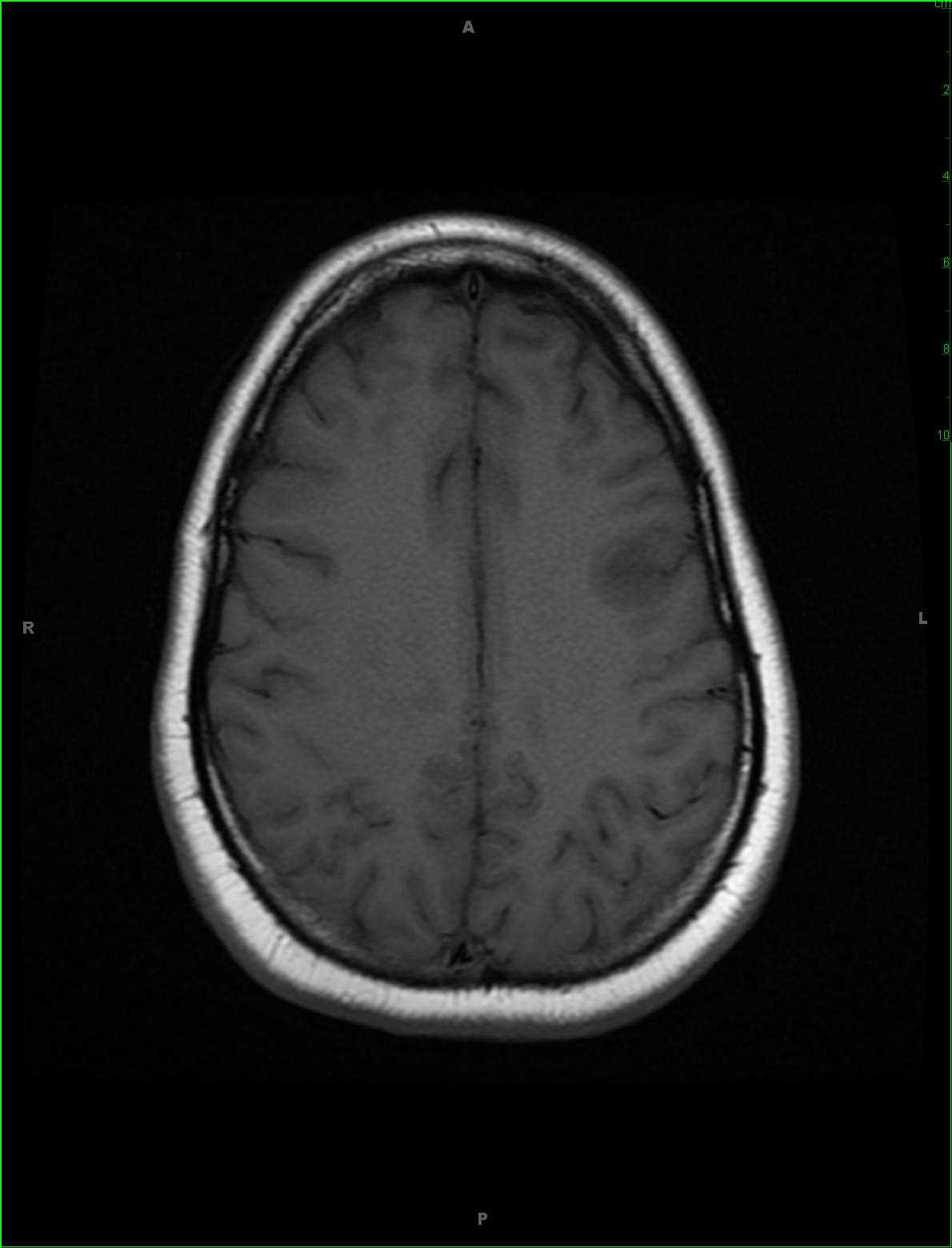
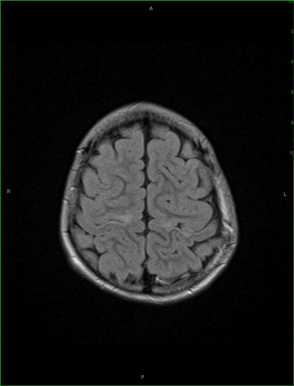
18-year-old female with history of prematurity and prior hypoxic ischemic insult. There is gliosis with volume loss and T2/FLAIR hyperintense signal along the corticospinal and thalamocortical tracts extending through the supratentorial white matter. Wallerian degeneration with cystic encephalomalacia is noted within the basal ganglia bilaterally with longitudinal T2/FLAIR hyperintense signal abnormality involving the corticospinal tracts as they descend through the cerebral peduncles. There is T1 hypointense signal at these sites compatible with gliotic change. The imaging findings are compatible with the stated clinical history of hypoxic ischemic injury. Hypoxic ischemic cerebral injury can occur at any age although etiology is age-dependent. In older children, the cause is usually drowning and asphyxiation. In adults, and causes more commonly the result of cardiac arrest or cerebrovascular disease. Severe acute ischemic injury primarily affects gray matter structures such as the basal ganglia, thalami, cerebral cortex, cerebellar and hippocampi. Neurologic injury is caused by hypoxia or interruption of blood flow.
Related videos to the case